


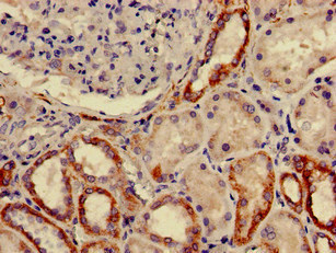
Immunohistochemical staining of human epididymis shows strong cytoplasmic positivity in glandular cells.
Immunohistochemical staining of human epididymis shows strong cytoplasmic positivity in glandular cells.
Anti-FURIN Antibody
HPA067869
ApplicationsImmunoHistoChemistry
Product group Antibodies
ReactivityHuman
TargetFURIN
Overview
- SupplierAtlas Antibodies
- Product NameAnti-FURIN Antibody
- Delivery Days Customer4
- ApplicationsImmunoHistoChemistry
- CertificationResearch Use Only
- ClonalityPolyclonal
- ConjugateUnconjugated
- Gene ID5045
- Target nameFURIN
- Target descriptionfurin, paired basic amino acid cleaving enzyme
- Target synonymsFUR, PACE, PCSK3, SPC1, furin, FES upstream region, dibasic processing enzyme, furin, membrane associated receptor protein, paired basic amino acid residue-cleaving enzyme, proprotein convertase subtilisin/kexin 3, proprotein convertase subtilisin/kexin type 3, subtilisin-like proprotein convertase 1
- HostRabbit
- IsotypeIgG
- Protein IDP09958
- Protein NameFurin
- Scientific DescriptionRecombinant Protein Epitope Signature Tag (PrEST) antigen sequence
- ReactivityHuman
- Storage Instruction-20°C,2°C to 8°C
- UNSPSC41116161
Datasheet
MSDS
Related products
Product group Antibodies
FURIN AntibodyCSB-PA009068LA01HU
ApplicationsELISA, ImmunoHistoChemistry
ReactivityHuman
TargetFURIN
- SizePrice
Product group Antibodies
Anti-Furin AntibodyA326245
ApplicationsWestern Blot, ELISA
ReactivityHuman
- SizePrice
Product group Antibodies

FURIN AntibodyLS-C748390
ApplicationsWestern Blot
ReactivityHuman, Mouse
TargetFURIN
- SizePrice
Product group Antibodies

Goat anti-FURIN / PCSK3EB08896
ApplicationsImmunoFluorescence, ELISA
ReactivityHuman, Mouse, Rat
TargetFURIN
- SizePrice
Product group Antibodies
Furin (FUR) Polyclonal AntibodyCAU24208
ApplicationsImmunoPrecipitation, Western Blot, ImmunoCytoChemistry, ImmunoHistoChemistry
ReactivityMouse, Porcine, Rat
TargetFURIN
- SizePrice

Product group Antibodies
Anti-Furin Antibody Picoband(r)PB9182-CARRIER-FREE
ApplicationsWestern Blot
ReactivityHuman
TargetFURIN
- SizePrice
Product group Antibodies
Furin Recombinant AntibodyBSM-54283R
ApplicationsImmunoFluorescence, Western Blot, ImmunoHistoChemistry, ImmunoHistoChemistry Frozen, ImmunoHistoChemistry Paraffin
ReactivityHuman, Mouse, Rat
TargetFURIN
- SizePrice
Product group Antibodies
Furin antibody [C3], C-termGTX109674
ApplicationsELISA
ReactivityHuman
TargetFURIN
- SizePrice