


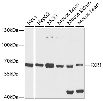
Figure 1. Western blot analysis of FXR1 using anti-FXR1 antibody (M03308). Electrophoresis was performed on a 5-20% SDS-PAGE gel at 70V (Stacking gel) / 90V (Resolving gel) for 2-3 hours. The sample well of each lane was loaded with 30 ug of sample under reducing conditions. Lane 1: human MCF-7 whole cell lysates, Lane 2: human 293T whole cell lysates, Lane 3: human Hela whole cell lysates, Lane 4: human HepG2 whole cell lysates, Lane 5: mouse heart tissue lysates. After electrophoresis, proteins were transferred to a nitrocellulose membrane at 150 mA for 50-90 minutes. Blocked the membrane with 5% non-fat milk/TBS for 1.5 hour at RT. The membrane was incubated with rabbit anti-FXR1 antigen affinity purified monoclonal antibody (Catalog # M03308) at 1:500 overnight at 4°C, then washed with TBS-0.1%Tween 3 times with 5 minutes each and probed with a goat anti-rabbit IgG-HRP secondary antibody at a dilution of 1:5000 for 1.5 hour at RT. The signal is developed using an Enhanced Chemiluminescent detection (ECL) kit (Catalog # EK1002) with Tanon 5200 system. A specific band was detected for FXR1 at approximately 70-80 kDa. The expected band size for FXR1 is at 70 kDa.
. FXR1 was detected in a paraffin-embedded section of mouse brain tissue. Heat mediated antigen retrieval was performed in EDTA buffer (pH 8.0, epitope retrieval solution). The tissue section was blocked with 10% goat serum. The tissue section was then incubated with 1:50 rabbit anti-FXR1 Antibody (M03308) overnight at 4°C. Peroxidase Conjugated Goat Anti-rabbit IgG was used as secondary antibody and incubated for 30 minutes at 37°C. The tissue section was developed using HRP Conjugated Rabbit IgG Super Vision Assay Kit (Catalog # SV0002) with DAB as the chromogen.")
. FXR1 was detected in a paraffin-embedded section of rat brain tissue. Heat mediated antigen retrieval was performed in EDTA buffer (pH 8.0, epitope retrieval solution). The tissue section was blocked with 10% goat serum. The tissue section was then incubated with 1:50 rabbit anti-FXR1 Antibody (M03308) overnight at 4°C. Peroxidase Conjugated Goat Anti-rabbit IgG was used as secondary antibody and incubated for 30 minutes at 37°C. The tissue section was developed using HRP Conjugated Rabbit IgG Super Vision Assay Kit (Catalog # SV0002) with DAB as the chromogen.")

Figure 1. Western blot analysis of FXR1 using anti-FXR1 antibody (M03308). Electrophoresis was performed on a 5-20% SDS-PAGE gel at 70V (Stacking gel) / 90V (Resolving gel) for 2-3 hours. The sample well of each lane was loaded with 30 ug of sample under reducing conditions. Lane 1: human MCF-7 whole cell lysates, Lane 2: human 293T whole cell lysates, Lane 3: human Hela whole cell lysates, Lane 4: human HepG2 whole cell lysates, Lane 5: mouse heart tissue lysates. After electrophoresis, proteins were transferred to a nitrocellulose membrane at 150 mA for 50-90 minutes. Blocked the membrane with 5% non-fat milk/TBS for 1.5 hour at RT. The membrane was incubated with rabbit anti-FXR1 antigen affinity purified monoclonal antibody (Catalog # M03308) at 1:500 overnight at 4°C, then washed with TBS-0.1%Tween 3 times with 5 minutes each and probed with a goat anti-rabbit IgG-HRP secondary antibody at a dilution of 1:5000 for 1.5 hour at RT. The signal is developed using an Enhanced Chemiluminescent detection (ECL) kit (Catalog # EK1002) with Tanon 5200 system. A specific band was detected for FXR1 at approximately 70-80 kDa. The expected band size for FXR1 is at 70 kDa.
Anti-FXR1 Rabbit Monoclonal Antibody
M03308
ApplicationsFlow Cytometry, ImmunoFluorescence, Western Blot, ImmunoCytoChemistry, ImmunoHistoChemistry
Product group Antibodies
ReactivityHuman, Mouse, Rat
TargetFXR1
Overview
- SupplierBoster Bio
- Product NameAnti-FXR1 Rabbit Monoclonal Antibody
- Delivery Days Customer9
- ApplicationsFlow Cytometry, ImmunoFluorescence, Western Blot, ImmunoCytoChemistry, ImmunoHistoChemistry
- CertificationResearch Use Only
- ClonalityMonoclonal
- Clone ID25F02
- Gene ID8087
- Target nameFXR1
- Target descriptionFMR1 autosomal homolog 1
- Target synonymsCMYO9A, CMYO9B, CMYP9A, CMYP9B, FXR1P, MYOPMIL, MYORIBF, RNA-binding protein FXR1, FMR1 autosomal protein-like protein 1
- HostRabbit
- IsotypeIgG
- Protein IDP51114
- Protein NameRNA-binding protein FXR1
- Scientific DescriptionBoster Bio Anti-FXR1 Rabbit Monoclonal Antibody catalog # M03308. Tested in WB, IHC, ICC/IF, Flow Cytometry applications. This antibody reacts with Human, Mouse, Rat.
- ReactivityHuman, Mouse, Rat
- Storage Instruction-20°C
- UNSPSC12352203
Related products
Product group Antibodies
FXR1 Recombinant AntibodyBSM-62314R
ApplicationsFlow Cytometry, ImmunoFluorescence, Western Blot, ImmunoCytoChemistry, ImmunoHistoChemistry, ImmunoHistoChemistry Frozen, ImmunoHistoChemistry Paraffin
ReactivityHuman, Mouse, Rat
TargetFXR1
- SizePrice
Product group Antibodies
FXR1 Polyclonal AntibodyCAC14724
ApplicationsImmunoFluorescence, Western Blot, ELISA, ImmunoHistoChemistry
ReactivityMouse
TargetFXR1
- SizePrice
Product group Antibodies
Anti-FXR1 AntibodyA15059
ApplicationsWestern Blot
ReactivityHuman, Mouse, Rat
- SizePrice
Product group Antibodies
Anti-FXR1 Antibody144-05942
ApplicationsImmunoFluorescence, Western Blot
ReactivityHuman, Mouse, Rat
TargetFXR1
- SizePrice
Product group Antibodies

Goat anti-FXR1 AntibodyEB07361
ApplicationsFlow Cytometry, ImmunoFluorescence, Western Blot, ELISA
ReactivityBovine, Canine, Human, Mouse, Rat
TargetFXR1
- SizePrice
Product group Antibodies
References
FXR1 antibodyGTX113867
ApplicationsImmunoFluorescence, ImmunoPrecipitation, Western Blot, ImmunoCytoChemistry, ImmunoHistoChemistry, ImmunoHistoChemistry Paraffin
ReactivityHuman, Mouse, Rat, Zebra Fish
TargetFXR1
- SizePrice
Product group Antibodies

FXR1 AntibodyLS-C334404
ApplicationsWestern Blot
ReactivityHuman, Mouse, Rat
TargetFXR1
- SizePrice

Product group Antibodies
Anti-FXR1 AntibodyHPA055475
ApplicationsImmunoCytoChemistry
ReactivityHuman
TargetFXR1
- SizePrice