

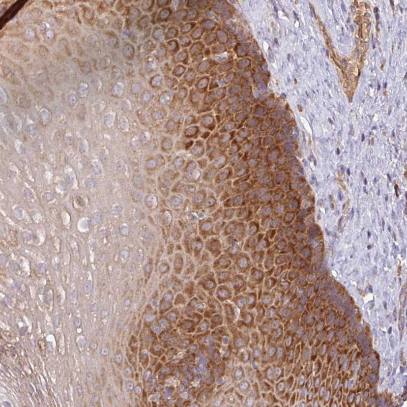
Immunohistochemical staining of human esophagus shows strong cytoplasmic positivity in squamous epithelial cells.
Immunohistochemical staining of human esophagus shows strong cytoplasmic positivity in squamous epithelial cells.
Anti-GALP Antibody
HPA053938
ApplicationsImmunoHistoChemistry
Product group Antibodies
ReactivityHuman
TargetGALP
Overview
- SupplierAtlas Antibodies
- Product NameAnti-GALP Antibody
- Delivery Days Customer4
- ApplicationsImmunoHistoChemistry
- CertificationResearch Use Only
- ClonalityPolyclonal
- ConjugateUnconjugated
- Gene ID85569
- Target nameGALP
- Target descriptiongalanin like peptide
- Target synonymsGAL, galanin-like peptide, alarin, gal-like peptide
- HostRabbit
- IsotypeIgG
- Protein IDQ9UBC7
- Protein NameGalanin-like peptide
- Scientific DescriptionRecombinant Protein Epitope Signature Tag (PrEST) antigen sequence
- ReactivityHuman
- Storage Instruction-20°C,2°C to 8°C
- UNSPSC41116161
Datasheet
MSDS
Related products
Product group Antibodies
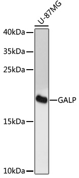
Anti-GALP AntibodyA88724
ApplicationsWestern Blot
ReactivityHuman, Mouse, Rat
- SizePrice

Product group Antibodies
Anti-GALP Antibody Picoband(r)A04786-1-CARRIER-FREE
ApplicationsWestern Blot, ELISA
ReactivityHuman, Mouse, Rat
TargetGALP
- SizePrice
Product group Antibodies
Anti-GALP Antibody130-10416
ApplicationsWestern Blot, ELISA
TargetGALP
- SizePrice
Product group Antibodies

ApplicationsWestern Blot, ELISA
ReactivityHuman
TargetGALP
- SizePrice
