

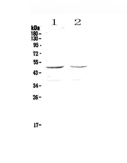
Figure 1. Western blot analysis of GALT using anti-GALT antibody (A01460-1). Electrophoresis was performed on a 5-20% SDS-PAGE gel at 70V (Stacking gel) / 90V (Resolving gel) for 2-3 hours. The sample well of each lane was loaded with 50ug of sample under reducing conditions. Lane 1: human HepG2 cell lysates, Lane 2: human MCF-7 cell lysates. After Electrophoresis, proteins were transferred to a Nitrocellulose membrane at 150mA for 50-90 minutes. Blocked the membrane with 5% Non-fat Milk/ TBS for 1.5 hour at RT. The membrane was incubated with rabbit anti-GALT antigen affinity purified polyclonal antibody (Catalog # A01460-1) at 0.5 microg/mL overnight at 4°C, then washed with TBS-0.1%Tween 3 times with 5 minutes each and probed with a goat anti-rabbit IgG-HRP secondary antibody at a dilution of 1:10000 for 1.5 hour at RT. The signal is developed using an Enhanced Chemiluminescent detection (ECL) kit (Catalog # EK1002) with Tanon 5200 system. A specific band was detected for GALT at approximately 48KD. The expected band size for GALT is at 43KD.
Figure 1. Western blot analysis of GALT using anti-GALT antibody (A01460-1). Electrophoresis was performed on a 5-20% SDS-PAGE gel at 70V (Stacking gel) / 90V (Resolving gel) for 2-3 hours. The sample well of each lane was loaded with 50ug of sample under reducing conditions. Lane 1: human HepG2 cell lysates, Lane 2: human MCF-7 cell lysates. After Electrophoresis, proteins were transferred to a Nitrocellulose membrane at 150mA for 50-90 minutes. Blocked the membrane with 5% Non-fat Milk/ TBS for 1.5 hour at RT. The membrane was incubated with rabbit anti-GALT antigen affinity purified polyclonal antibody (Catalog # A01460-1) at 0.5 microg/mL overnight at 4°C, then washed with TBS-0.1%Tween 3 times with 5 minutes each and probed with a goat anti-rabbit IgG-HRP secondary antibody at a dilution of 1:10000 for 1.5 hour at RT. The signal is developed using an Enhanced Chemiluminescent detection (ECL) kit (Catalog # EK1002) with Tanon 5200 system. A specific band was detected for GALT at approximately 48KD. The expected band size for GALT is at 43KD.
Anti-GALT Antibody Picoband(r)
A01460-1-FITC
ApplicationsFlow Cytometry, ImmunoPrecipitation, Western Blot, ImmunoHistoChemistry
Product group Antibodies
ReactivityHuman, Rat
TargetGALT
Overview
- SupplierBoster Bio
- Product NameAnti-GALT Antibody Picoband(r)
- Delivery Days Customer9
- ApplicationsFlow Cytometry, ImmunoPrecipitation, Western Blot, ImmunoHistoChemistry
- CertificationResearch Use Only
- ClonalityPolyclonal
- Concentration500 ug/ml
- ConjugateFITC
- Gene ID2592
- Target nameGALT
- Target descriptiongalactose-1-phosphate uridylyltransferase
- Target synonymsgalactose-1-phosphate uridylyltransferase, UDP-glucose--hexose-1-phosphate uridylyltransferase, gal-1-P uridylyltransferase, galactose-1-phosphate uridyl transferase
- HostRabbit
- IsotypeIgG
- Protein IDP07902
- Protein NameGalactose-1-phosphate uridylyltransferase
- Scientific DescriptionBoster Bio Anti-GALT Antibody Picoband® catalog # A01460-1. Tested in ELISA, Flow Cytometry, IP, IHC, WB applications. This antibody reacts with Human, Rat. The brand Picoband indicates this is a premium antibody that guarantees superior quality, high affinity, and strong signals with minimal background in Western blot applications. Only our best-performing antibodies are designated as Picoband, ensuring unmatched performance.
- ReactivityHuman, Rat
- Storage Instruction-20°C,2°C to 8°C
- UNSPSC12352203
Related products
Product group Antibodies
Galt Polyclonal AntibodyCAC08457
ApplicationsImmunoFluorescence, ImmunoPrecipitation, Western Blot, ELISA, ImmunoHistoChemistry
ReactivityMouse, Rat
TargetGALT
- SizePrice
Product group Antibodies
GALT Polyclonal AntibodyBS-3984R
ApplicationsImmunoFluorescence, Western Blot, ELISA, ImmunoCytoChemistry, ImmunoHistoChemistry, ImmunoHistoChemistry Frozen, ImmunoHistoChemistry Paraffin
ReactivityBovine, Human, Mouse, Rat
TargetGALT
- SizePrice
Product group Antibodies
Anti-GALT AntibodyA31271
ApplicationsWestern Blot, ImmunoHistoChemistry
ReactivityHuman, Mouse, Rat
- SizePrice
Product group Antibodies
Anti-GALT Antibody144-06292
ApplicationsWestern Blot, ImmunoHistoChemistry
ReactivityHuman, Mouse, Rat
TargetGALT
- SizePrice
Product group Antibodies
GALT antibody [N3C3]GTX113702
ApplicationsWestern Blot
ReactivityHuman
TargetGALT
- SizePrice
Product group Antibodies

GALT AntibodyLS-C405004
ApplicationsWestern Blot, ELISA, ImmunoHistoChemistry
ReactivityHuman, Mouse, Rat
TargetGALT
- SizePrice

Product group Antibodies
Anti-GALT AntibodyHPA004868
ApplicationsWestern Blot, ImmunoHistoChemistry
ReactivityHuman
TargetGALT
- SizePrice
Product group Antibodies
GALT AntibodyCSB-PA009226LA01HU
ApplicationsImmunoFluorescence, ImmunoPrecipitation, Western Blot, ELISA, ImmunoHistoChemistry
ReactivityHuman, Mouse, Rat
TargetGALT
- SizePrice
Product group Antibodies
Anti-GALT Antibody Picoband(r)A01460-1-CARRIER-FREE
ApplicationsFlow Cytometry, ImmunoPrecipitation, Western Blot, ImmunoHistoChemistry
ReactivityHuman, Rat
TargetGALT
- SizePrice