

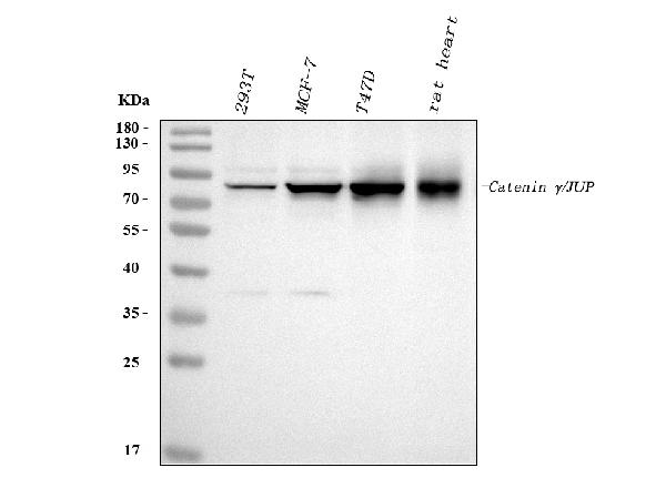
Figure 1. Western blot analysis of gamma Catenin using anti-gamma Catenin antibody (PB9185). Electrophoresis was performed on a 5-20% SDS-PAGE gel at 70V (Stacking gel) / 90V (Resolving gel) for 2-3 hours. The sample well of each lane was loaded with 30 ug of sample under reducing conditions. Lane 1: human 293T whole cell lysates, Lane 2: human MCF-7 whole cell lysates, Lane 3: human T47D whole cell lysates, Lane 4: rat heart tissue lysates. After electrophoresis, proteins were transferred to a nitrocellulose membrane at 150 mA for 50-90 minutes. Blocked the membrane with 5% non-fat milk/TBS for 1.5 hour at RT. The membrane was incubated with rabbit anti-gamma Catenin antigen affinity purified polyclonal antibody (Catalog # PB9185) at 0.5 microg/mL overnight at 4°C, then washed with TBS-0.1%Tween 3 times with 5 minutes each and probed with a goat anti-rabbit IgG-HRP secondary antibody at a dilution of 1:5000 for 1.5 hour at RT. The signal is developed using an Enhanced Chemiluminescent detection (ECL) kit (Catalog # EK1002) with Tanon 5200 system. A specific band was detected for gamma Catenin at approximately 82 kDa. The expected band size for gamma Catenin is at 82 kDa.
. gamma Catenin was detected in paraffin-embedded section of human intestinal cancer tissue. Heat mediated antigen retrieval was performed in citrate buffer (pH6, epitope retrieval solution) for 20 mins. The tissue section was blocked with 10% goat serum. The tissue section was then incubated with 1microg/ml rabbit anti-gamma Catenin Antibody (PB9185) overnight at 4°C. Biotinylated goat anti-rabbit IgG was used as secondary antibody and incubated for 30 minutes at 37°C. The tissue section was developed using Strepavidin-Biotin-Complex (SABC)(Catalog # SA1022) with DAB as the chromogen.")
. gamma Catenin was detected in paraffin-embedded section of mouse intestine tissue. Heat mediated antigen retrieval was performed in citrate buffer (pH6, epitope retrieval solution) for 20 mins. The tissue section was blocked with 10% goat serum. The tissue section was then incubated with 1microg/ml rabbit anti-gamma Catenin Antibody (PB9185) overnight at 4°C. Biotinylated goat anti-rabbit IgG was used as secondary antibody and incubated for 30 minutes at 37°C. The tissue section was developed using Strepavidin-Biotin-Complex (SABC)(Catalog # SA1022) with DAB as the chromogen.")
. gamma Catenin was detected in paraffin-embedded section of rat intestine tissue. Heat mediated antigen retrieval was performed in citrate buffer (pH6, epitope retrieval solution) for 20 mins. The tissue section was blocked with 10% goat serum. The tissue section was then incubated with 1microg/ml rabbit anti-gamma Catenin Antibody (PB9185) overnight at 4°C. Biotinylated goat anti-rabbit IgG was used as secondary antibody and incubated for 30 minutes at 37°C. The tissue section was developed using Strepavidin-Biotin-Complex (SABC)(Catalog # SA1022) with DAB as the chromogen.")
. gamma Catenin was detected in frozen section of rat intestine tissue. The tissue section was blocked with 10% goat serum. The tissue section was then incubated with 1microg/ml rabbit anti-gamma Catenin Antibody (PB9185) overnight at 4°C. Biotinylated goat anti-rabbit IgG was used as secondary antibody and incubated for 30 minutes at 37°C. The tissue section was developed using Strepavidin-Biotin-Complex (SABC)(Catalog # SA1022) with DAB as the chromogen.")
. gamma Catenin was detected in immunocytochemical section of human MCF-7 cell. Enzyme antigen retrieval was performed using IHC enzyme antigen retrieval reagent (AR0022) for 15 mins. The cells were blocked with 10% goat serum. And then incubated with 1microg/ml rabbit anti-gamma Catenin Antibody (PB9185) overnight at 4°C. Biotinylated goat anti-rabbit IgG was used as secondary antibody and incubated for 30 minutes at 37°C. The section was developed using Strepavidin-Biotin-Complex (SABC)(Catalog # SA1022) with DAB as the chromogen.")
. gamma Catenin was detected in immunocytochemical section of A431 cells. Enzyme antigen retrieval was performed using IHC enzyme antigen retrieval reagent (AR0022) for 15 mins. The cells were blocked with 10% goat serum. And then incubated with 2microg/mL rabbit anti-gamma Catenin Antibody (PB9185) overnight at 4°C. DyLight®488 Conjugated Goat Anti-Rabbit IgG (BA1127) was used as secondary antibody at 1:100 dilution and incubated for 30 minutes at 37°C. The section was counterstained with DAPI. Visualize using a fluorescence microscope and filter sets appropriate for the label used.")
. Overlay histogram showing A431 cells stained with PB9185 (Blue line). To facilitate intracellular staining, cells were fixed with 4% paraformaldehyde and permeabilized with permeabilization buffer. The cells were blocked with 10% normal goat serum. And then incubated with rabbit anti-gamma Catenin Antibody (PB9185,1microg/1x106 cells) for 30 min at 20°C. DyLight®488 conjugated goat anti-rabbit IgG (BA1127, 5-10microg/1x106 cells) was used as secondary antibody for 30 minutes at 20°C. Isotype control antibody (Green line) was rabbit IgG (1microg/1x106) used under the same conditions. Unlabelled sample without incubation with primary antibody and secondary antibody (Red line) was used as a blank control.")
Figure 1. Western blot analysis of gamma Catenin using anti-gamma Catenin antibody (PB9185). Electrophoresis was performed on a 5-20% SDS-PAGE gel at 70V (Stacking gel) / 90V (Resolving gel) for 2-3 hours. The sample well of each lane was loaded with 30 ug of sample under reducing conditions. Lane 1: human 293T whole cell lysates, Lane 2: human MCF-7 whole cell lysates, Lane 3: human T47D whole cell lysates, Lane 4: rat heart tissue lysates. After electrophoresis, proteins were transferred to a nitrocellulose membrane at 150 mA for 50-90 minutes. Blocked the membrane with 5% non-fat milk/TBS for 1.5 hour at RT. The membrane was incubated with rabbit anti-gamma Catenin antigen affinity purified polyclonal antibody (Catalog # PB9185) at 0.5 microg/mL overnight at 4°C, then washed with TBS-0.1%Tween 3 times with 5 minutes each and probed with a goat anti-rabbit IgG-HRP secondary antibody at a dilution of 1:5000 for 1.5 hour at RT. The signal is developed using an Enhanced Chemiluminescent detection (ECL) kit (Catalog # EK1002) with Tanon 5200 system. A specific band was detected for gamma Catenin at approximately 82 kDa. The expected band size for gamma Catenin is at 82 kDa.
Anti-gamma Catenin/JUP Antibody Picoband(r)
PB9185-FITC
ApplicationsFlow Cytometry, ImmunoFluorescence, Western Blot, ImmunoCytoChemistry, ImmunoHistoChemistry, ImmunoHistoChemistry Frozen
Product group Antibodies
ReactivityHuman, Mouse, Rat
TargetJUP
Overview
- SupplierBoster Bio
- Product NameAnti-gamma Catenin/JUP Antibody Picoband(r)
- Delivery Days Customer9
- Application Supplier NoteWB: The detection limit for gamma Catenin is approximately 0.25ng/lane under reducing conditions. Tested Species: In-house tested species with positive results. Predicted Species: Species predicted to be fit for the product based on sequence similarities. By Heat: Boiling the paraffin sections in 10mM citrate buffer, pH6.0, for 20mins is required for the staining of formalin/paraffin sections. Other applications have not been tested. Optimal dilutions should be determined by end users.
- ApplicationsFlow Cytometry, ImmunoFluorescence, Western Blot, ImmunoCytoChemistry, ImmunoHistoChemistry, ImmunoHistoChemistry Frozen
- CertificationResearch Use Only
- ClonalityPolyclonal
- Concentration500 ug/ml
- ConjugateFITC
- Gene ID3728
- Target nameJUP
- Target descriptionjunction plakoglobin
- Target synonymsCTNNG, DP3, DPIII, PDGB, PG, PKGB, junction plakoglobin, catenin (cadherin-associated protein), gamma 80kDa, desmoplakin III, desmoplakin-3, desmosomal protein 3
- HostRabbit
- IsotypeIgG
- Protein IDP14923
- Protein NameJunction plakoglobin
- Scientific DescriptionBoster Bio Anti-gamma Catenin/JUP Antibody Picoband® catalog # PB9185. Tested in Flow Cytometry, IF, IHC, IHC-F, ICC, WB applications. This antibody reacts with Human, Mouse, Rat. The brand Picoband indicates this is a premium antibody that guarantees superior quality, high affinity, and strong signals with minimal background in Western blot applications. Only our best-performing antibodies are designated as Picoband, ensuring unmatched performance.
- ReactivityHuman, Mouse, Rat
- Storage Instruction-20°C,2°C to 8°C
- UNSPSC12352203
Related products
Product group Antibodies
Anti-JUP Antibody144-00963
ApplicationsImmunoFluorescence, ImmunoPrecipitation, Western Blot
ReactivityHuman, Mouse, Rat
TargetJUP
- SizePrice
Product group Antibodies
JUP Polyclonal AntibodyCAC14134
ApplicationsWestern Blot, ELISA, ImmunoHistoChemistry
TargetJUP
- SizePrice
Product group Antibodies
gamma Catenin Recombinant Antibody, Biotin ConjugatedBSM-61391R-BIOTIN
ApplicationsWestern Blot, ImmunoHistoChemistry, ImmunoHistoChemistry Frozen, ImmunoHistoChemistry Paraffin
ReactivityCanine, Human, Mouse, Rat
TargetJUP
- SizePrice
Product group Antibodies
JUP AntibodyCSB-PA001293
ApplicationsImmunoFluorescence, Western Blot, ELISA, ImmunoHistoChemistry
ReactivityHuman, Mouse, Rat
TargetJUP
- SizePrice
Product group Antibodies
ApplicationsWestern Blot, ELISA, ImmunoHistoChemistry
ReactivityHuman, Mouse, Rat
- SizePrice

Product group Antibodies
Anti-JUP AntibodyHPA032047
ApplicationsWestern Blot, ImmunoHistoChemistry
ReactivityHuman
TargetJUP
- SizePrice
Product group Antibodies

ApplicationsWestern Blot, ELISA
ReactivityHuman, Mouse, Rat
TargetJUP
- SizePrice
Product group Antibodies

JUP/CTNNG/Junction Plakoglobin AntibodyLS-C401790
ApplicationsWestern Blot, ELISA, ImmunoHistoChemistry
ReactivityHuman, Mouse, Rat
TargetJUP
- SizePrice