


Anti-GAP43 Antibody
A85292
ApplicationsImmunoFluorescence, Western Blot, ImmunoCytoChemistry, ImmunoHistoChemistry
Product group Antibodies
ReactivityBovine, Equine, Human, Mouse, Porcine, Rat
Overview
- SupplierAntibodies.com
- Product NameAnti-GAP43 Antibody
- Delivery Days Customer7
- ApplicationsImmunoFluorescence, Western Blot, ImmunoCytoChemistry, ImmunoHistoChemistry
- CertificationResearch Use Only
- ClonalityPolyclonal
- ConjugateUnconjugated
- HostChicken
- IsotypeIgY
- Scientific DescriptionChicken polyclonal antibody to GAP43.
- ReactivityBovine, Equine, Human, Mouse, Porcine, Rat
- UNSPSC12352203
Related products
Product group Antibodies
Anti-GAP43 Antibody144-06376
ApplicationsWestern Blot
ReactivityHuman, Mouse, Rat
TargetGAP43
- SizePrice

Product group Antibodies
Anti-GAP43 Antibody Picoband(r)A01868-CARRIER-FREE
ApplicationsFlow Cytometry, ImmunoFluorescence, Western Blot, ImmunoHistoChemistry
ReactivityHuman, Mouse, Rat
TargetGAP43
- SizePrice

Product group Antibodies
Anti-GAP43 AntibodyAMAB91664
ApplicationsImmunoHistoChemistry
ReactivityHuman
TargetGAP43
- SizePrice
Product group Antibodies
ApplicationsWestern Blot, ELISA, ImmunoCytoChemistry, ImmunoHistoChemistry, ImmunoHistoChemistry Frozen, ImmunoHistoChemistry Paraffin
ReactivityPorcine, Rat
TargetGAP43
- SizePrice
Product group Antibodies
GAP43 Monoclonal AntibodyCSB-MA080173
ApplicationsWestern Blot, ELISA, ImmunoHistoChemistry
ReactivityHuman, Mouse, Rat
TargetGAP43
- SizePrice
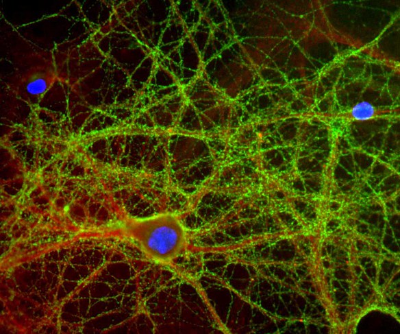
![GAP43 antibody detects GAP43 protein by immunofluorescent analysis. Sample: DIV9 rat E18 primary cortical neurons were fixed in 4% paraformaldehyde at RT for 15 min. Green: GAP43 protein stained by GAP43 antibody (GTX101535) diluted at 1:500. Red: beta Tubulin 3/ Tuj1, stained by beta Tubulin 3/ Tuj1 antibody [GT886] (GTX631830) diluted at 1:500. Blue: Fluoroshield with DAPI (GTX30920).](https://www.genetex.com/upload/website/prouct_img/normal/GTX101535/GTX101535_40016_20170705_IFA_R_w_23060100_168.webp)
Product group Antibodies
GAP43 antibodyGTX101535
ApplicationsImmunoFluorescence, Western Blot, ImmunoCytoChemistry
ReactivityHuman, Mouse, Rat
TargetGAP43
- SizePrice
Product group Antibodies
ApplicationsFlow Cytometry, ImmunoFluorescence, ELISA, ImmunoCytoChemistry, ImmunoHistoChemistry, ImmunoHistoChemistry Frozen, ImmunoHistoChemistry Paraffin
ReactivityCanine, Human, Mouse, Rat
TargetGAP43
- SizePrice
Product group Antibodies
Anti-GAP43 AntibodyCAB16857
ApplicationsWestern Blot, ELISA, ImmunoHistoChemistry, ImmunoHistoChemistry Paraffin
ReactivityMouse
- SizePrice
Product group Antibodies

GAP43 Antibody (clone 6F2)LS-C772675
ApplicationsWestern Blot, ImmunoHistoChemistry, ImmunoHistoChemistry Paraffin
ReactivityHuman, Mouse, Rat
TargetGAP43
- SizePrice