


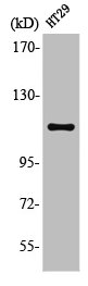
Figure 1. Western blot analysis of GEF-H1/ARHGEF2 using anti-GEF-H1/ARHGEF2 antibody (A02572-3). Electrophoresis was performed on a 5-20% SDS-PAGE gel at 70V (Stacking gel) / 90V (Resolving gel) for 2-3 hours. The sample well of each lane was loaded with 30 ug of sample under reducing conditions. Lane 1: human 293T whole cell lysates, Lane 2: human Hela whole cell lysates, Lane 3: human PC-3 whole cell lysates, Lane 4: rat brain tissue lysates, Lane 5: rat C6 whole cell lysates, Lane 6: mouse brain tissue lysates, Lane 7: mouse Raw264.7 whole cell lysates. After electrophoresis, proteins were transferred to a nitrocellulose membrane at 150 mA for 50-90 minutes. Blocked the membrane with 5% non-fat milk/TBS for 1.5 hour at RT. The membrane was incubated with rabbit anti-GEF-H1/ARHGEF2 antigen affinity purified polyclonal antibody (Catalog # A02572-3) at 0.5 microg/mL overnight at 4°C, then washed with TBS-0.1%Tween 3 times with 5 minutes each and probed with a goat anti-rabbit IgG-HRP secondary antibody at a dilution of 1:5000 for 1.5 hour at RT. The signal is developed using an Enhanced Chemiluminescent detection (ECL) kit (Catalog # EK1002) with Tanon 5200 system. A specific band was detected for GEF-H1/ARHGEF2 at approximately 112 kDa. The expected band size for GEF-H1/ARHGEF2 is at 112 kDa.
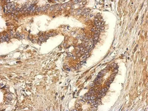
. GEF-H1/ARHGEF2 was detected in a paraffin-embedded section of human colon cancer tissue. Heat mediated antigen retrieval was performed in EDTA buffer (pH 8.0, epitope retrieval solution). The tissue section was blocked with 10% goat serum. The tissue section was then incubated with 2 microg/ml rabbit anti-GEF-H1/ARHGEF2 Antibody (A02572-3) overnight at 4°C. Peroxidase Conjugated Goat Anti-rabbit IgG was used as secondary antibody and incubated for 30 minutes at 37°C. The tissue section was developed using HRP Conjugated Rabbit IgG Super Vision Assay Kit (Catalog # SV0002) with DAB as the chromogen.")
. GEF-H1/ARHGEF2 was detected in a paraffin-embedded section of human ovarian cancer tissue. Heat mediated antigen retrieval was performed in EDTA buffer (pH 8.0, epitope retrieval solution). The tissue section was blocked with 10% goat serum. The tissue section was then incubated with 2 microg/ml rabbit anti-GEF-H1/ARHGEF2 Antibody (A02572-3) overnight at 4°C. Peroxidase Conjugated Goat Anti-rabbit IgG was used as secondary antibody and incubated for 30 minutes at 37°C. The tissue section was developed using HRP Conjugated Rabbit IgG Super Vision Assay Kit (Catalog # SV0002) with DAB as the chromogen.")
. GEF-H1/ARHGEF2 was detected in a paraffin-embedded section of human ovary cancer tissue. Heat mediated antigen retrieval was performed in EDTA buffer (pH 8.0, epitope retrieval solution). The tissue section was blocked with 10% goat serum. The tissue section was then incubated with 2 microg/ml rabbit anti-GEF-H1/ARHGEF2 Antibody (A02572-3) overnight at 4°C. Peroxidase Conjugated Goat Anti-rabbit IgG was used as secondary antibody and incubated for 30 minutes at 37°C. The tissue section was developed using HRP Conjugated Rabbit IgG Super Vision Assay Kit (Catalog # SV0002) with DAB as the chromogen.")
. Overlay histogram showing PC-3 cells stained with A02572-3 (Blue line). To facilitate intracellular staining, cells were fixed with 4% paraformaldehyde and permeabilized with permeabilization buffer. The cells were blocked with 10% normal goat serum. And then incubated with rabbit anti-GEF-H1/ARHGEF2 Antibody (A02572-3, 1 microg/1x106 cells) for 30 min at 20°C. DyLight®488 conjugated goat anti-rabbit IgG (BA1127, 5-10 microg/1x106 cells) was used as secondary antibody for 30 minutes at 20°C. Isotype control antibody (Green line) was rabbit IgG (1 microg/1x106) used under the same conditions. Unlabelled sample without incubation with primary antibody and secondary antibody (Red line) was used as a blank control.")
Figure 1. Western blot analysis of GEF-H1/ARHGEF2 using anti-GEF-H1/ARHGEF2 antibody (A02572-3). Electrophoresis was performed on a 5-20% SDS-PAGE gel at 70V (Stacking gel) / 90V (Resolving gel) for 2-3 hours. The sample well of each lane was loaded with 30 ug of sample under reducing conditions. Lane 1: human 293T whole cell lysates, Lane 2: human Hela whole cell lysates, Lane 3: human PC-3 whole cell lysates, Lane 4: rat brain tissue lysates, Lane 5: rat C6 whole cell lysates, Lane 6: mouse brain tissue lysates, Lane 7: mouse Raw264.7 whole cell lysates. After electrophoresis, proteins were transferred to a nitrocellulose membrane at 150 mA for 50-90 minutes. Blocked the membrane with 5% non-fat milk/TBS for 1.5 hour at RT. The membrane was incubated with rabbit anti-GEF-H1/ARHGEF2 antigen affinity purified polyclonal antibody (Catalog # A02572-3) at 0.5 microg/mL overnight at 4°C, then washed with TBS-0.1%Tween 3 times with 5 minutes each and probed with a goat anti-rabbit IgG-HRP secondary antibody at a dilution of 1:5000 for 1.5 hour at RT. The signal is developed using an Enhanced Chemiluminescent detection (ECL) kit (Catalog # EK1002) with Tanon 5200 system. A specific band was detected for GEF-H1/ARHGEF2 at approximately 112 kDa. The expected band size for GEF-H1/ARHGEF2 is at 112 kDa.
Anti-GEF-H1/ARHGEF2 Antibody Picoband(r)
A02572-3-CARRIER-FREE
ApplicationsFlow Cytometry, Western Blot, ELISA, ImmunoHistoChemistry
Product group Antibodies
ReactivityHuman, Mouse, Rat
TargetARHGEF2
Overview
- SupplierBoster Bio
- Product NameAnti-GEF-H1/ARHGEF2 Antibody Picoband(r)
- Delivery Days Customer9
- ApplicationsFlow Cytometry, Western Blot, ELISA, ImmunoHistoChemistry
- CertificationResearch Use Only
- ClonalityPolyclonal
- Concentration500 ug/ml
- Gene ID9181
- Target nameARHGEF2
- Target descriptionRho/Rac guanine nucleotide exchange factor 2
- Target synonymsGEF, GEF-H1, GEFH1, LFP40, Lfc, NEDMHM, P40, rho guanine nucleotide exchange factor 2, Rho/Rac guanine nucleotide exchange factor (GEF) 2, guanine nucleotide exchange factor H1, microtubule-regulated Rho-GEF, proliferating cell nucleolar antigen p40
- HostRabbit
- IsotypeIgG
- Protein IDQ92974
- Protein NameRho guanine nucleotide exchange factor 2
- Scientific DescriptionBoster Bio Anti-GEF-H1/ARHGEF2 Antibody Picoband® catalog # A02572-3. Tested in WB, IHC, Flow Cytometry, ELISA applications. This antibody reacts with Human, Mouse, Rat. The brand Picoband indicates this is a premium antibody that guarantees superior quality, high affinity, and strong signals with minimal background in Western blot applications. Only our best-performing antibodies are designated as Picoband, ensuring unmatched performance.
- ReactivityHuman, Mouse, Rat
- Storage Instruction-20°C,2°C to 8°C
- UNSPSC12352203
Related products
Product group Antibodies
Anti-ARHGEF2 AntibodyA100186
ApplicationsWestern Blot, ELISA
ReactivityHuman
- SizePrice

Product group Antibodies
GEF H1 Recombinant AntibodyBSM-54369R
ApplicationsFlow Cytometry, ImmunoFluorescence, Western Blot, ImmunoCytoChemistry, ImmunoHistoChemistry, ImmunoHistoChemistry Paraffin
ReactivityHuman, Mouse, Rat
TargetARHGEF2
- SizePrice
Product group Antibodies
ARHGEF2 AntibodyCSB-PA003148
ApplicationsWestern Blot, ELISA
ReactivityHuman, Mouse, Rat
TargetARHGEF2
- SizePrice
Product group Antibodies

ARHGEF2 / GEF-H1 AntibodyLS-C404553
ApplicationsELISA, ImmunoHistoChemistry
ReactivityHuman, Mouse, Rat
TargetARHGEF2
- SizePrice

Product group Antibodies
Anti-ARHGEF2 AntibodyHPA043437
ApplicationsImmunoHistoChemistry
ReactivityHuman
TargetARHGEF2
- SizePrice
Product group Antibodies
GEF-H1 antibodyGTX125893
ApplicationsImmunoFluorescence, ImmunoPrecipitation, Western Blot, ImmunoCytoChemistry, ImmunoHistoChemistry, ImmunoHistoChemistry Frozen, ImmunoHistoChemistry Paraffin
ReactivityHuman, Mouse, Rat
TargetARHGEF2
- SizePrice