

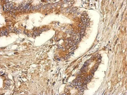
Immunohistochemical analysis of paraffin-embedded human gastric cancer, using GEF-H1(GTX125893) antibody at 1:500 dilution.
Antigen Retrieval: Trilogy? (EDTA based, pH 8.0) buffer, 15min
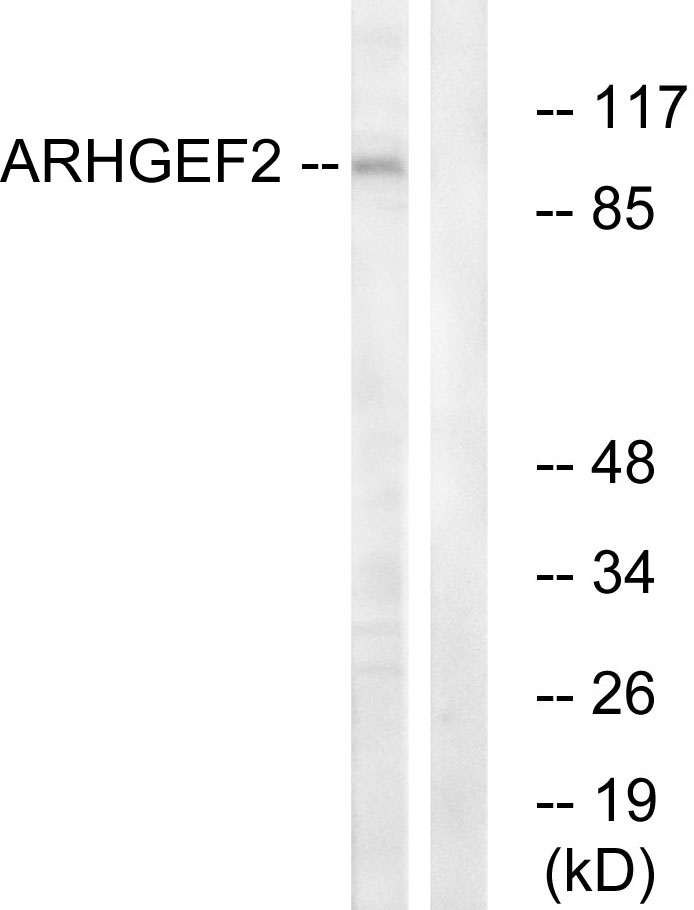
 were separated by 7.5% SDS-PAGE, and the membrane was blotted with GEF-H1 antibody (GTX125893) diluted at 1:500. The HRP-conjugated anti-rabbit IgG antibody (GTX213110-01) was used to detect the primary antibody.")
 and transfected (+) 293T whole cell extracts (30 μg) were separated by 7.5% SDS-PAGE, and the membrane was blotted with GEF-H1 antibody (GTX125893) diluted at 1:4000. The HRP-conjugated anti-rabbit IgG antibody (GTX213110-01) was used to detect the primary antibody.")
 A: hMSC-3A6 B: siRNA of ARHGEF2/GEF-H1 8% SDS PAGE GTX125893 diluted at 1:1000 The HRP-conjugated anti-rabbit IgG antibody (GTX213110-01) was used to detect the primary antibody.")
![GEF-H1 antibody detects GEF-H1 protein by immunohistochemical analysis. Sample: Frozen-sectioned mouse mouse cerebellum. Green: GEF-H1 stained by GEF-H1 antibody (GTX125893) diluted at 1:250. Red: NF-H, stained by NF-H antibody [GT114] (GTX634289) diluted at 1:500. Blue: Fluoroshield with DAPI (GTX30920).
Antigen Retrieval: Citrate buffer, pH 6.0, 10 min](https://www.genetex.com/upload/website/prouct_img/normal/GTX125893/GTX125893_41570_20180530_IHC-Fr_M_2_w_23060522_798.webp "GEF-H1 antibody detects GEF-H1 protein by immunohistochemical analysis. Sample: Frozen-sectioned mouse mouse cerebellum. Green: GEF-H1 stained by GEF-H1 antibody (GTX125893) diluted at 1:250. Red: NF-H, stained by NF-H antibody [GT114] (GTX634289) diluted at 1:500. Blue: Fluoroshield with DAPI (GTX30920).
Antigen Retrieval: Citrate buffer, pH 6.0, 10 min")
 dilution: 1:1000 The HRP-conjugated anti-rabbit IgG antibody (GTX213110-01) was used to detect the primary antibody.")
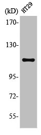
. Western blot analysis was performed using GEF-H1 antibody (GTX125893). EasyBlot anti-Rabbit IgG (GTX221666-01) was used as a secondary reagent.")
 dilution: 1:500.
Antigen Retrieval: Trilogy? (EDTA based, pH 8.0) buffer, 15min")
 dilution: 1:500.
Antigen Retrieval: Trilogy? (EDTA based, pH 8.0) buffer, 15min")
 diluted at 1:500. Blue: Hoechst 33342 staining. Scale bar= 10 μm.")
Immunohistochemical analysis of paraffin-embedded human gastric cancer, using GEF-H1(GTX125893) antibody at 1:500 dilution.
Antigen Retrieval: Trilogy? (EDTA based, pH 8.0) buffer, 15min
GEF-H1 antibody
GTX125893
ApplicationsImmunoFluorescence, ImmunoPrecipitation, Western Blot, ImmunoCytoChemistry, ImmunoHistoChemistry, ImmunoHistoChemistry Frozen, ImmunoHistoChemistry Paraffin
Product group Antibodies
ReactivityHuman, Mouse, Rat
TargetARHGEF2
Overview
- SupplierGeneTex
- Product NameGEF-H1 antibody
- Delivery Days Customer9
- Application Supplier NoteWB: 1:500-1:3000. ICC/IF: 1:100-1:1000. IHC-P: 1:100-1:1000. IHC-Fr: 1:100-1:1000. IP: 1:100-1:500. *Optimal dilutions/concentrations should be determined by the researcher.Not tested in other applications.
- ApplicationsImmunoFluorescence, ImmunoPrecipitation, Western Blot, ImmunoCytoChemistry, ImmunoHistoChemistry, ImmunoHistoChemistry Frozen, ImmunoHistoChemistry Paraffin
- CertificationResearch Use Only
- ClonalityPolyclonal
- Concentration0.71 mg/ml
- ConjugateUnconjugated
- Gene ID9181
- Target nameARHGEF2
- Target descriptionRho/Rac guanine nucleotide exchange factor 2
- Target synonymsGEF, GEF-H1, GEFH1, LFP40, Lfc, NEDMHM, P40, rho guanine nucleotide exchange factor 2, Rho/Rac guanine nucleotide exchange factor (GEF) 2, guanine nucleotide exchange factor H1, microtubule-regulated Rho-GEF, proliferating cell nucleolar antigen p40
- HostRabbit
- IsotypeIgG
- Protein IDQ92974
- Protein NameRho guanine nucleotide exchange factor 2
- Scientific DescriptionRho GTPases play a fundamental role in numerous cellular processes that are initiated by extracellular stimuli that work through G protein coupled receptors. The encoded protein may form complex with G proteins and stimulate rho-dependent signals. [provided by RefSeq]
- ReactivityHuman, Mouse, Rat
- Storage Instruction-20°C or -80°C,2°C to 8°C
- UNSPSC41116161
Datasheet
Related products
Product group Antibodies
Anti-ARHGEF2 AntibodyA100186
ApplicationsWestern Blot, ELISA
ReactivityHuman
- SizePrice

Product group Antibodies
Anti-GEF-H1/ARHGEF2 Antibody Picoband(r)A02572-3-CARRIER-FREE
ApplicationsFlow Cytometry, Western Blot, ELISA, ImmunoHistoChemistry
ReactivityHuman, Mouse, Rat
TargetARHGEF2
- SizePrice

Product group Antibodies
GEF H1 Recombinant AntibodyBSM-54369R
ApplicationsFlow Cytometry, ImmunoFluorescence, Western Blot, ImmunoCytoChemistry, ImmunoHistoChemistry, ImmunoHistoChemistry Paraffin
ReactivityHuman, Mouse, Rat
TargetARHGEF2
- SizePrice
Product group Antibodies
ARHGEF2 AntibodyCSB-PA003148
ApplicationsWestern Blot, ELISA
ReactivityHuman, Mouse, Rat
TargetARHGEF2
- SizePrice
Product group Antibodies

ARHGEF2 / GEF-H1 AntibodyLS-C404553
ApplicationsELISA, ImmunoHistoChemistry
ReactivityHuman, Mouse, Rat
TargetARHGEF2
- SizePrice

Product group Antibodies
Anti-ARHGEF2 AntibodyHPA043437
ApplicationsImmunoHistoChemistry
ReactivityHuman
TargetARHGEF2
- SizePrice
![Various whole cell extracts (30 μg) were separated by 7.5% SDS-PAGE, and the membrane was blotted with GEF-H1 antibody [HL3470] (GTX641355) diluted at 1:1000. The HRP-conjugated anti-rabbit IgG antibody (GTX213110-01) was used to detect the primary antibody.](https://www.genetex.com/upload/website/prouct_img/normal/GTX641355/GTX641355_T-45600_20241122_WB_R_24112622_975.webp)
Product group Antibodies
GEF-H1 antibody [HL3470]GTX641355
ApplicationsImmunoFluorescence, Western Blot, ImmunoCytoChemistry, ImmunoHistoChemistry, ImmunoHistoChemistry Paraffin
ReactivityHuman, Mouse, Rat
TargetARHGEF2
- SizePrice
Product group Antibodies
GEF-H1 antibody [B4/7]GTX54457
ApplicationsImmunoFluorescence, ImmunoPrecipitation, Western Blot, ImmunoCytoChemistry, ImmunoHistoChemistry, ImmunoHistoChemistry Frozen
ReactivityCanine, Human
TargetARHGEF2
- SizePrice
![GEF-H1 antibody [GT336] detects GEF-H1 protein by Western blot analysis. A. 30 μg 293T whole cell lysate/extract B. 30 μg A431 whole cell lysate/extract C. 30 μg HeLa whole cell lysate/extract 7.5 % SDS-PAGE GEF-H1 antibody [GT336] (GTX629398) dilution: 1:500](https://www.genetex.com/upload/website/prouct_img/normal/GTX629398/GTX629398_41323_WB_w_23061202_354.webp)
Product group Antibodies
GEF-H1 antibody [GT336]GTX629398
ApplicationsImmunoFluorescence, Western Blot, ImmunoCytoChemistry
ReactivityHuman, Mouse, Rat
TargetARHGEF2
- SizePrice