


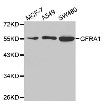
Figure 1. Western blot analysis of GFRA1 using anti-GFRA1 antibody (PB9202). Electrophoresis was performed on a 5-20% SDS-PAGE gel at 70V (Stacking gel) / 90V (Resolving gel) for 2-3 hours. Lane 1: recombinant human GFRA1 protein 0.5 ng. After electrophoresis, proteins were transferred to a nitrocellulose membrane at 150 mA for 50-90 minutes. Blocked the membrane with 5% non-fat milk/TBS for 1.5 hour at RT. The membrane was incubated with rabbit anti-GFRA1 antigen affinity purified polyclonal antibody (Catalog # PB9202) at 0.5 microg/mL overnight at 4°C, then washed with TBS-0.1%Tween 3 times with 5 minutes each and probed with a goat anti-rabbit IgG-HRP secondary antibody at a dilution of 1:5000 for 1.5 hour at RT. The signal is developed using an Enhanced Chemiluminescent detection (ECL) kit (Catalog # EK1002) with Tanon 5200 system. A specific band was detected for GFRA1 at approximately 39 kDa. The expected band size for GFRA1 is at 39 kDa.
. Electrophoresis was performed on a 5-20% SDS-PAGE gel at 70V (Stacking gel) / 90V (Resolving gel) for 2-3 hours. The sample well of each lane was loaded with 30 ug of sample under reducing conditions. Lane 1: human placenta tissue lysates, After electrophoresis, proteins were transferred to a nitrocellulose membrane at 150 mA for 50-90 minutes. Blocked the membrane with 5% non-fat milk/TBS for 1.5 hour at RT. The membrane was incubated with rabbit anti-GFRA1 antigen affinity purified polyclonal antibody (Catalog # PB9202) at 0.5 microg/mL overnight at 4°C, then washed with TBS-0.1%Tween 3 times with 5 minutes each and probed with a goat anti-rabbit IgG-HRP secondary antibody at a dilution of 1:5000 for 1.5 hour at RT. The signal is developed using an Enhanced Chemiluminescent detection (ECL) kit (Catalog # EK1002) with Tanon 5200 system. A specific band was detected for GFRA1 at approximately 51 kDa. The expected band size for GFRA1 is at 51 kDa.")
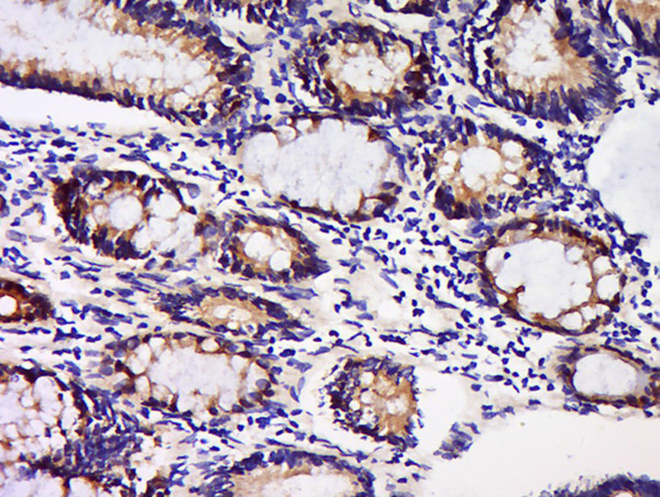
. GFRA1 was detected in a paraffin-embedded section of human lung cancer tissue. Heat mediated antigen retrieval was performed in EDTA buffer (pH 8.0, epitope retrieval solution). The tissue section was blocked with 10% goat serum. The tissue section was then incubated with 1 microg/ml rabbit anti-GFRA1 Antibody (PB9202) overnight at 4°C. Biotinylated goat anti-rabbit IgG was used as secondary antibody and incubated for 30 minutes at 37°C. The tissue section was developed using Strepavidin-Biotin-Complex (SABC) (Catalog # SA1022) with DAB as the chromogen.")
Figure 1. Western blot analysis of GFRA1 using anti-GFRA1 antibody (PB9202). Electrophoresis was performed on a 5-20% SDS-PAGE gel at 70V (Stacking gel) / 90V (Resolving gel) for 2-3 hours. Lane 1: recombinant human GFRA1 protein 0.5 ng. After electrophoresis, proteins were transferred to a nitrocellulose membrane at 150 mA for 50-90 minutes. Blocked the membrane with 5% non-fat milk/TBS for 1.5 hour at RT. The membrane was incubated with rabbit anti-GFRA1 antigen affinity purified polyclonal antibody (Catalog # PB9202) at 0.5 microg/mL overnight at 4°C, then washed with TBS-0.1%Tween 3 times with 5 minutes each and probed with a goat anti-rabbit IgG-HRP secondary antibody at a dilution of 1:5000 for 1.5 hour at RT. The signal is developed using an Enhanced Chemiluminescent detection (ECL) kit (Catalog # EK1002) with Tanon 5200 system. A specific band was detected for GFRA1 at approximately 39 kDa. The expected band size for GFRA1 is at 39 kDa.
Anti-GFRA1 Picoband Antibody
PB9202
ApplicationsWestern Blot, ImmunoHistoChemistry
Product group Antibodies
ReactivityHuman
TargetGFRA1
Overview
- SupplierBoster Bio
- Product NameAnti-GFRA1 Picoband Antibody
- Delivery Days Customer9
- Application Supplier NoteWB: The detection limit for GFRA1 is approximately 0.2ng/lane under reducing conditions. Tested Species: In-house tested species with positive results. By Heat: Boiling the paraffin sections in 10mM citrate buffer, pH6.0, for 20mins is required for the staining of formalin/paraffin sections. Other applications have not been tested. Optimal dilutions should be determined by end users.
- ApplicationsWestern Blot, ImmunoHistoChemistry
- Applications SupplierIHP, WB, IHC
- CertificationResearch Use Only
- ClonalityPolyclonal
- Concentration500 ug/ml
- Gene ID2674
- Target nameGFRA1
- Target descriptionGDNF family receptor alpha 1
- Target synonymsGDNFR, GDNFR-alpha-1, GDNFRA, GFR-ALPHA-1, GFRalpha-1, RET1L, RETL1, RHDA4, TRNR1, GDNF family receptor alpha-1, GDNF receptor alpha 1d, GDNF receptor alpha 1e, GPI-linked anchor protein, Glial cell line-derived neurotrophic factor receptor alpha, PI-linked cell-surface accessory protein, RET ligand 1, TGF-beta-related neurotrophic factor receptor 1
- HostRabbit
- IsotypeIgG
- Protein IDP56159
- Protein NameGDNF family receptor alpha-1
- Scientific DescriptionBoster Bio Anti-GDNF Receptor alpha 1/GFRA1 Antibody Picoband® catalog # PB9202. Tested in IHC, WB applications. This antibody reacts with Human. The brand Picoband indicates this is a premium antibody that guarantees superior quality, high affinity, and strong signals with minimal background in Western blot applications. Only our best-performing antibodies are designated as Picoband, ensuring unmatched performance.
- ReactivityHuman
- Reactivity SupplierHuman
- Storage Instruction-20°C,2°C to 8°C
- UNSPSC12352203
References
- Wang J, Liu L, Li Z, et al. JMJD3 regulate H3K27me3 modification via interacting directly with TET1 to affect spermatogonia self-renewal and proliferation. BMC Genomics. 2024,25(1):225. doi: 10.1186/s12864-024-10120-9Read this paper
- Liu L, Wang J, Wang S, et al. Epigenetic Regulation of TET1-SP1 During Spermatogonia Self-Renewal and Proliferation. Front Physiol. 2022,13:843825. doi: 10.3389/fphys.2022.843825Read this paper
- Zhu WQ, Cai NN, Jiang Y, et al. Survivable potential of germ cells after trehalose cryopreservation of bovine testicular tissues. Cryobiology. 2021,101:105-114. doi: 10.1016/j.cryobiol.2021.05.001Read this paper
- Jiang Y, Zhu WQ, Zhu XC, et al. Cryopreservation of calf testicular tissues with knockout serum replacement. Cryobiology. 2020,92:255-257. doi: 10.1016/j.cryobiol.2020.01.010Read this paper
- Cai H, Tang B, Wu JY, et al. Enrichment and in vitro features of the putative gonocytes from cryopreserved testicular tissue of neonatal bulls. Andrology. 2016,4(6):1150-1158. doi: 10.1111/andr.12229Read this paper
Datasheet
MSDS
Related products
Product group Antibodies
Gfra1 Polyclonal AntibodyCAC07821
ApplicationsImmunoFluorescence, Western Blot, ELISA, ImmunoHistoChemistry
ReactivityMouse
TargetGFRA1
- SizePrice
Product group Antibodies
References
GDNFRA Polyclonal AntibodyBS-0201R
ApplicationsFlow Cytometry, ImmunoFluorescence, Western Blot, ELISA, ImmunoCytoChemistry, ImmunoHistoChemistry, ImmunoHistoChemistry Frozen, ImmunoHistoChemistry Paraffin
ReactivityBovine, Canine, Equine, Human, Mouse, Porcine, Rat
TargetGFRA1
- SizePrice
Product group Antibodies
Anti-GFRA1 Antibody144-65897
ApplicationsImmunoFluorescence, Western Blot
ReactivityHuman, Mouse, Rat
TargetGFRA1
- SizePrice
Product group Antibodies

GFR alpha 1 AntibodyABX013085
ApplicationsELISA, ImmunoHistoChemistry
- SizePrice
Product group Antibodies
Anti-GFRA1 AntibodyA35275
ApplicationsImmunoFluorescence, Western Blot, ImmunoHistoChemistry
ReactivityHuman, Mouse, Rat
- SizePrice
Product group Antibodies
GDNF Receptor alpha 1 antibodyGTX11115
ApplicationsWestern Blot, ELISA, ImmunoHistoChemistry, ImmunoHistoChemistry Paraffin
ReactivityHuman, Mouse, Rat
TargetGFRA1
- SizePrice
Product group Antibodies

GFRA1 / GFR Alpha AntibodyLS-C832593
ApplicationsWestern Blot, ELISA, ImmunoHistoChemistry
ReactivityHuman, Mouse, Rat
TargetGFRA1
- SizePrice

Product group Antibodies
Anti-GFRA1 AntibodyHPA043829
ApplicationsImmunoCytoChemistry, ImmunoHistoChemistry
ReactivityHuman
TargetGFRA1
- SizePrice
Product group Antibodies
GFRA1 AntibodyCSB-PA008804
ApplicationsELISA, ImmunoHistoChemistry
ReactivityHuman, Mouse, Rat
TargetGFRA1
- SizePrice