


Figure 1. Western blot analysis of Giantin/GOLGB1 using anti-Giantin/GOLGB1 antibody (A08226-1). Electrophoresis was performed on a 5-20% SDS-PAGE gel at 70V (Stacking gel) / 90V (Resolving gel) for 2-3 hours. The sample well of each lane was loaded with 30 ug of sample under reducing conditions. Lane 1: human Hela whole cell lysates. After electrophoresis, proteins were transferred to a nitrocellulose membrane at 150 mA for 50-90 minutes. Blocked the membrane with 5% non-fat milk/TBS for 1.5 hour at RT. The membrane was incubated with rabbit anti-Giantin/GOLGB1 antigen affinity purified polyclonal antibody (Catalog # A08226-1) at 0.5 microg/mL overnight at 4°C, then washed with TBS-0.1%Tween 3 times with 5 minutes each and probed with a goat anti-rabbit IgG-HRP secondary antibody at a dilution of 1:5000 for 1.5 hour at RT. The signal is developed using an Enhanced Chemiluminescent detection (ECL) kit (Catalog # EK1002) with Tanon 5200 system. A specific band was detected for Giantin/GOLGB1 at approximately 376 kDa. The expected band size for Giantin/GOLGB1 is at 376 kDa.
. Giantin/GOLGB1 was detected in a paraffin-embedded section of human colonic adenocarcinoma tissue. Heat mediated antigen retrieval was performed in EDTA buffer (pH 8.0, epitope retrieval solution). The tissue section was blocked with 10% goat serum. The tissue section was then incubated with 2 microg/ml rabbit anti-Giantin/GOLGB1 Antibody (A08226-1) overnight at 4°C. Biotinylated goat anti-rabbit IgG was used as secondary antibody and incubated for 30 minutes at 37°C. The tissue section was developed using Strepavidin-Biotin-Complex (SABC) (Catalog # SA1022) with DAB as the chromogen.")
. Giantin/GOLGB1 was detected in a paraffin-embedded section of human esophageal squamous carcinoma tissue. Heat mediated antigen retrieval was performed in EDTA buffer (pH 8.0, epitope retrieval solution). The tissue section was blocked with 10% goat serum. The tissue section was then incubated with 2 microg/ml rabbit anti-Giantin/GOLGB1 Antibody (A08226-1) overnight at 4°C. Biotinylated goat anti-rabbit IgG was used as secondary antibody and incubated for 30 minutes at 37°C. The tissue section was developed using Strepavidin-Biotin-Complex (SABC) (Catalog # SA1022) with DAB as the chromogen.")
. Giantin/GOLGB1 was detected in a paraffin-embedded section of human gall bladder adenosquamous carcinoma tissue. Heat mediated antigen retrieval was performed in EDTA buffer (pH 8.0, epitope retrieval solution). The tissue section was blocked with 10% goat serum. The tissue section was then incubated with 2 microg/ml rabbit anti-Giantin/GOLGB1 Antibody (A08226-1) overnight at 4°C. Biotinylated goat anti-rabbit IgG was used as secondary antibody and incubated for 30 minutes at 37°C. The tissue section was developed using Strepavidin-Biotin-Complex (SABC) (Catalog # SA1022) with DAB as the chromogen.")
. Giantin/GOLGB1 was detected in a paraffin-embedded section of human placenta tissue. Heat mediated antigen retrieval was performed in EDTA buffer (pH 8.0, epitope retrieval solution). The tissue section was blocked with 10% goat serum. The tissue section was then incubated with 2 microg/ml rabbit anti-Giantin/GOLGB1 Antibody (A08226-1) overnight at 4°C. Biotinylated goat anti-rabbit IgG was used as secondary antibody and incubated for 30 minutes at 37°C. The tissue section was developed using Strepavidin-Biotin-Complex (SABC) (Catalog # SA1022) with DAB as the chromogen.")
. Giantin/GOLGB1 was detected in a paraffin-embedded section of human appendiceal adenocarcinoma tissue. Heat mediated antigen retrieval was performed in EDTA buffer (pH 8.0, epitope retrieval solution). The tissue section was blocked with 10% goat serum. The tissue section was then incubated with 2 microg/ml rabbit anti-Giantin/GOLGB1 Antibody (A08226-1) overnight at 4°C. Biotinylated goat anti-rabbit IgG was used as secondary antibody and incubated for 30 minutes at 37°C. The tissue section was developed using Strepavidin-Biotin-Complex (SABC) (Catalog # SA1022) with DAB as the chromogen.")
. Giantin/GOLGB1 was detected in a paraffin-embedded section of human breast cancer tissue. Heat mediated antigen retrieval was performed in EDTA buffer (pH 8.0, epitope retrieval solution). The tissue section was blocked with 10% goat serum. The tissue section was then incubated with 2 microg/ml rabbit anti-Giantin/GOLGB1 Antibody (A08226-1) overnight at 4°C. Biotinylated goat anti-rabbit IgG was used as secondary antibody and incubated for 30 minutes at 37°C. The tissue section was developed using Strepavidin-Biotin-Complex (SABC) (Catalog # SA1022) with DAB as the chromogen.")
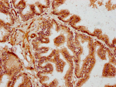
. Giantin/GOLGB1 was detected in a paraffin-embedded section of human gastric adenocarcinoma tissue. Heat mediated antigen retrieval was performed in EDTA buffer (pH 8.0, epitope retrieval solution). The tissue section was blocked with 10% goat serum. The tissue section was then incubated with 2 microg/ml rabbit anti-Giantin/GOLGB1 Antibody (A08226-1) overnight at 4°C. Biotinylated goat anti-rabbit IgG was used as secondary antibody and incubated for 30 minutes at 37°C. The tissue section was developed using Strepavidin-Biotin-Complex (SABC) (Catalog # SA1022) with DAB as the chromogen.")
. Giantin/GOLGB1 was detected in an immunocytochemical section of RT4 cells. Enzyme antigen retrieval was performed using IHC enzyme antigen retrieval reagent (AR0022) for 15 mins. The cells were blocked with 10% goat serum. And then incubated with 5 microg/mL rabbit anti-Giantin/GOLGB1 Antibody (A08226-1) overnight at 4°C. DyLight®488 Conjugated Goat Anti-Rabbit IgG (BA1127) was used as secondary antibody at 1:100 dilution and incubated for 30 minutes at 37°C. The section was counterstained with DAPI. Visualize using a fluorescence microscope and filter sets appropriate for the label used.")
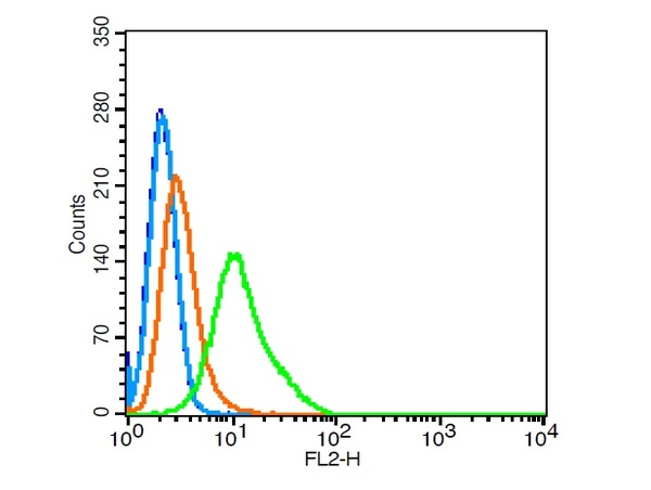
. Overlay histogram showing HepG2 cells stained with A08226-1 (Blue line). To facilitate intracellular staining, cells were fixed with 4% paraformaldehyde and permeabilized with permeabilization buffer. The cells were blocked with 10% normal goat serum. And then incubated with rabbit anti-Giantin/GOLGB1 Antibody (A08226-1, 1 microg/1x106 cells) for 30 min at 20°C. DyLight®488 conjugated goat anti-rabbit IgG (BA1127, 5-10 microg/1x106 cells) was used as secondary antibody for 30 minutes at 20°C. Isotype control antibody (Green line) was rabbit IgG (1 microg/1x106) used under the same conditions. Unlabelled sample without incubation with primary antibody and secondary antibody (Red line) was used as a blank control.")
Figure 1. Western blot analysis of Giantin/GOLGB1 using anti-Giantin/GOLGB1 antibody (A08226-1). Electrophoresis was performed on a 5-20% SDS-PAGE gel at 70V (Stacking gel) / 90V (Resolving gel) for 2-3 hours. The sample well of each lane was loaded with 30 ug of sample under reducing conditions. Lane 1: human Hela whole cell lysates. After electrophoresis, proteins were transferred to a nitrocellulose membrane at 150 mA for 50-90 minutes. Blocked the membrane with 5% non-fat milk/TBS for 1.5 hour at RT. The membrane was incubated with rabbit anti-Giantin/GOLGB1 antigen affinity purified polyclonal antibody (Catalog # A08226-1) at 0.5 microg/mL overnight at 4°C, then washed with TBS-0.1%Tween 3 times with 5 minutes each and probed with a goat anti-rabbit IgG-HRP secondary antibody at a dilution of 1:5000 for 1.5 hour at RT. The signal is developed using an Enhanced Chemiluminescent detection (ECL) kit (Catalog # EK1002) with Tanon 5200 system. A specific band was detected for Giantin/GOLGB1 at approximately 376 kDa. The expected band size for Giantin/GOLGB1 is at 376 kDa.
Anti-Giantin/GOLGB1 Antibody Picoband(r)
A08226-1-CARRIER-FREE
ApplicationsFlow Cytometry, ImmunoFluorescence, Western Blot, ELISA, ImmunoCytoChemistry, ImmunoHistoChemistry
Product group Antibodies
ReactivityHuman
TargetGOLGB1
Overview
- SupplierBoster Bio
- Product NameAnti-Giantin/GOLGB1 Antibody Picoband(r)
- Delivery Days Customer9
- ApplicationsFlow Cytometry, ImmunoFluorescence, Western Blot, ELISA, ImmunoCytoChemistry, ImmunoHistoChemistry
- CertificationResearch Use Only
- ClonalityPolyclonal
- Concentration500 ug/ml
- Gene ID2804
- Target nameGOLGB1
- Target descriptiongolgin B1
- Target synonymsGCP, GCP372, GOLIM1, golgin subfamily B member 1, 372 kDa Golgi complex-associated protein, giantin, golgi autoantigen, golgin subfamily b, macrogolgin (with transmembrane signal), 1, golgi integral membrane protein 1, golgin B1, golgi integral membrane protein
- HostRabbit
- IsotypeIgG
- Protein IDQ14789
- Protein NameGolgin subfamily B member 1
- Scientific DescriptionBoster Bio Anti-Giantin/GOLGB1 Antibody Picoband® catalog # A08226-1. Tested in ELISA, Flow Cytometry, IF, IHC, ICC, WB applications. This antibody reacts with Human. The brand Picoband indicates this is a premium antibody that guarantees superior quality, high affinity, and strong signals with minimal background in Western blot applications. Only our best-performing antibodies are designated as Picoband, ensuring unmatched performance.
- ReactivityHuman
- Storage Instruction-20°C,2°C to 8°C
- UNSPSC12352203
Related products
Product group Antibodies
anti-Giantin, mAb (rec.) (TA10)AG-27B-0003
ApplicationsImmunoCytoChemistry
ReactivityHuman, Mouse
TargetGOLGB1
- SizePrice
Product group Antibodies
Anti-GOLGB1 Antibody101-10650
ApplicationsWestern Blot, ELISA
TargetGOLGB1
- SizePrice
Product group Antibodies
Giantin Polyclonal AntibodyBS-13356R
ApplicationsFlow Cytometry, ImmunoFluorescence, Western Blot, ELISA, ImmunoCytoChemistry, ImmunoHistoChemistry, ImmunoHistoChemistry Frozen, ImmunoHistoChemistry Paraffin
ReactivityCanine, Equine, Hamster, Human, Monkey, Mouse, Rabbit, Rat
TargetGOLGB1
- SizePrice
Product group Antibodies
GOLGB1 AntibodyCSB-PA622788LA01HU
ApplicationsImmunoFluorescence, ELISA, ImmunoHistoChemistry
ReactivityHuman
TargetGOLGB1
- SizePrice
Product group Antibodies

GOLGB1 / Giantin Antibody (aa425-475)LS-C290678
ApplicationsImmunoPrecipitation, Western Blot
ReactivityHuman
TargetGOLGB1
- SizePrice

Product group Antibodies
Anti-GOLGB1 AntibodyHPA011555
ApplicationsImmunoCytoChemistry, ImmunoHistoChemistry
ReactivityHuman
TargetGOLGB1
- SizePrice
![Giantin antibody [TA10]](https://www.genetex.com/upload/website/prouct_img/normal/GTX33924/GTX33924_20160831_Structure_w_23060801_967.webp)
Product group Antibodies
Giantin antibody [TA10]GTX33924
ApplicationsImmunoFluorescence, ImmunoCytoChemistry
ReactivityHuman, Mouse
TargetGOLGB1
- SizePrice