


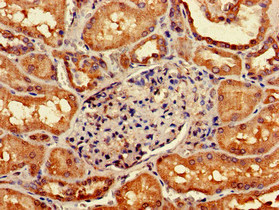
Immunohistochemical staining of human urinary bladder shows moderate cytoplasmic positivity in urothelial cells.
 shows similar pattern to independent antibody HPA061786 (B).")
Immunohistochemical staining of human urinary bladder shows moderate cytoplasmic positivity in urothelial cells.
Anti-GIPC1 Antibody
HPA043958
ApplicationsWestern Blot, ImmunoHistoChemistry
Product group Antibodies
ReactivityHuman
TargetGIPC1
Overview
- SupplierAtlas Antibodies
- Product NameAnti-GIPC1 Antibody
- Delivery Days Customer4
- ApplicationsWestern Blot, ImmunoHistoChemistry
- CertificationResearch Use Only
- ClonalityPolyclonal
- ConjugateUnconjugated
- Gene ID10755
- Target nameGIPC1
- Target descriptionGIPC PDZ domain containing family member 1
- Target synonymsC19orf3, GIPC, GLUT1CBP, Hs.6454, IIP-1, NIP, OPDM2, RGS19IP1, SEMCAP, SYNECTIIN, SYNECTIN, TIP-2, PDZ domain-containing protein GIPC1, GAIP C-terminus-interacting protein, GLUT1 C-terminal binding protein, IGF-1 receptor interacting protein 1, RGS-GAIP-interacting protein, RGS19-interacting protein 1, regulator of G-protein signalling 19 interacting protein 1, tax interaction protein 2
- HostRabbit
- IsotypeIgG
- Protein IDO14908
- Protein NamePDZ domain-containing protein GIPC1
- Scientific DescriptionRecombinant Protein Epitope Signature Tag (PrEST) antigen sequence
- ReactivityHuman
- Storage Instruction-20°C,2°C to 8°C
- UNSPSC41116161
Datasheet
MSDS
Related products
Product group Antibodies
GIPC1 AntibodyCSB-PA009435LA01HU
ApplicationsImmunoFluorescence, ELISA, ImmunoHistoChemistry
ReactivityHuman
TargetGIPC1
- SizePrice
Product group Antibodies
Anti-GIPC1 AntibodyA12992
ApplicationsWestern Blot
ReactivityHuman, Mouse, Rat
- SizePrice

Product group Antibodies
Anti-GIPC1 Antibody Picoband(r)A04969-4-CARRIER-FREE
ApplicationsFlow Cytometry, ImmunoFluorescence, Western Blot, ELISA, ImmunoCytoChemistry, ImmunoHistoChemistry
ReactivityHuman, Mouse, Rat
TargetGIPC1
- SizePrice
Product group Antibodies

GIPC1 / GIPC AntibodyLS-C830106
ApplicationsWestern Blot, ELISA
ReactivityHuman, Mouse, Rat
TargetGIPC1
- SizePrice
Product group Antibodies

Goat anti-GIPC1 / NIPEB06196
ApplicationsWestern Blot, ELISA
ReactivityBovine, Human, Mouse, Rat
TargetGIPC1
- SizePrice

Product group Antibodies
Anti-GIPC1 AntibodyHPA061786
ApplicationsWestern Blot
ReactivityHuman
TargetGIPC1
- SizePrice
Product group Antibodies
GIPC1 antibodyGTX106382
ApplicationsWestern Blot, ImmunoHistoChemistry, ImmunoHistoChemistry Paraffin
ReactivityHuman, Mouse
TargetGIPC1
- SizePrice
Product group Antibodies
Anti-GIPC1 Antibody144-10550
ApplicationsWestern Blot
ReactivityHuman, Mouse, Rat
TargetGIPC1
- SizePrice