


Figure 1. Western blot analysis of Tnfrsf18 using anti-Tnfrsf18 antibody (A03125-3). Electrophoresis was performed on a 5-20% SDS-PAGE gel at 70V (Stacking gel) / 90V (Resolving gel) for 2-3 hours. The sample well of each lane was loaded with 50ug of sample under reducing conditions. Lane 1: rat thymus tissue lysates, Lane 2: rat skeletal muscle tissue lysates, Lane 3: mouse skeletal muscle tissue lysates. After Electrophoresis, proteins were transferred to a Nitrocellulose membrane at 150mA for 50-90 minutes. Blocked the membrane with 5% Non-fat Milk/ TBS for 1.5 hour at RT. The membrane was incubated with rabbit anti-Tnfrsf18 antigen affinity purified polyclonal antibody (Catalog # A03125-3) at 0.5 microg/mL overnight at 4°C, then washed with TBS-0.1%Tween 3 times with 5 minutes each and probed with a goat anti-rabbit IgG-HRP secondary antibody at a dilution of 1:5000 for 1.5 hour at RT. The signal is developed using an Enhanced Chemiluminescent detection (ECL) kit (Catalog # EK1002) with Tanon 5200 system. A specific band was detected for Tnfrsf18 at approximately 30-35KD. The expected band size for Tnfrsf18 is at 26KD.
Figure 1. Western blot analysis of Tnfrsf18 using anti-Tnfrsf18 antibody (A03125-3). Electrophoresis was performed on a 5-20% SDS-PAGE gel at 70V (Stacking gel) / 90V (Resolving gel) for 2-3 hours. The sample well of each lane was loaded with 50ug of sample under reducing conditions. Lane 1: rat thymus tissue lysates, Lane 2: rat skeletal muscle tissue lysates, Lane 3: mouse skeletal muscle tissue lysates. After Electrophoresis, proteins were transferred to a Nitrocellulose membrane at 150mA for 50-90 minutes. Blocked the membrane with 5% Non-fat Milk/ TBS for 1.5 hour at RT. The membrane was incubated with rabbit anti-Tnfrsf18 antigen affinity purified polyclonal antibody (Catalog # A03125-3) at 0.5 microg/mL overnight at 4°C, then washed with TBS-0.1%Tween 3 times with 5 minutes each and probed with a goat anti-rabbit IgG-HRP secondary antibody at a dilution of 1:5000 for 1.5 hour at RT. The signal is developed using an Enhanced Chemiluminescent detection (ECL) kit (Catalog # EK1002) with Tanon 5200 system. A specific band was detected for Tnfrsf18 at approximately 30-35KD. The expected band size for Tnfrsf18 is at 26KD.
Anti-GITR/Tnfrsf18 Picoband(r) Antibody
A03125-3-CARRIER-FREE
ApplicationsWestern Blot, ELISA
Product group Antibodies
ReactivityMouse, Rat
TargetTnfrsf18
Overview
- SupplierBoster Bio
- Product NameAnti-GITR/Tnfrsf18 Picoband(r) Antibody
- Delivery Days Customer9
- ApplicationsWestern Blot, ELISA
- CertificationResearch Use Only
- ClonalityPolyclonal
- Concentration500 ug/ml
- Gene ID21936
- Target nameTnfrsf18
- Target descriptiontumor necrosis factor receptor superfamily, member 18
- Target synonymsAITR, Gitr, tumor necrosis factor receptor superfamily member 18, glucocorticoid-induced TNFR-related protein
- HostRabbit
- IsotypeIgG
- Protein IDO35714
- Protein NameTumor necrosis factor receptor superfamily member 18
- Scientific DescriptionBoster Bio Anti-GITR/Tnfrsf18 Picoband® Antibody catalog # A03125-3. Tested in ELISA, WB applications. This antibody reacts with Mouse, Rat. The brand Picoband indicates this is a premium antibody that guarantees superior quality, high affinity, and strong signals with minimal background in Western blot applications. Only our best-performing antibodies are designated as Picoband, ensuring unmatched performance.
- ReactivityMouse, Rat
- Storage Instruction-20°C,2°C to 8°C
- UNSPSC12352203
Related products
Product group Antibodies
anti-GITR (mouse), mAb (MGIT 02)AG-20A-0007
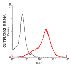
ApplicationsFlow Cytometry, ELISA
ReactivityMouse
TargetTnfrsf18
- SizePrice
Product group Antibodies
Anti-Mouse GITR Antibody (TNFRSF18)188-10843
ReactivityMouse
TargetTnfrsf18
- SizePrice
Product group Antibodies

Anti-GITR [DTA-1]AB01060-2.0-VXS
ApplicationsFlow Cytometry, ImmunoPrecipitation, Western Blot, Other Application
ReactivityMouse
TargetTnfrsf18
- SizePrice
Product group Antibodies
TNFRSF18 Polyclonal AntibodyBS-1173R
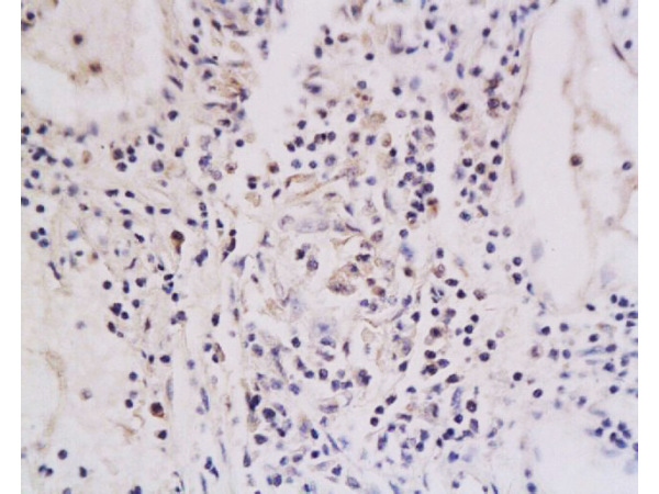
ApplicationsImmunoFluorescence, ELISA, ImmunoCytoChemistry, ImmunoHistoChemistry, ImmunoHistoChemistry Frozen, ImmunoHistoChemistry Paraffin
ReactivityHuman, Mouse, Rat
TargetTnfrsf18
- SizePrice
Product group Antibodies
GITR antibody [MAB0860]GTX52441
ApplicationsFlow Cytometry, Neutralisation/Blocking
ReactivityMouse
TargetTnfrsf18
- SizePrice