


Immunofluorescence staining of RH-30 cells using the Anti-GLI1 monoclonal antibody, showing specific staining in the nucleoplasm and cytosol in green. Microtubule- and nuclear probes are visualized in red and blue, respectively (where available).

Immunofluorescence staining of RH-30 cells using the Anti-GLI1 monoclonal antibody, showing specific staining in the nucleoplasm and cytosol in green. Microtubule- and nuclear probes are visualized in red and blue, respectively (where available).
Anti-GLI1 Antibody
AMAB91771
ApplicationsWestern Blot, ImmunoCytoChemistry
Product group Antibodies
ReactivityHuman
TargetGLI1
Overview
- SupplierAtlas Antibodies
- Product NameAnti-GLI1 Antibody
- Delivery Days Customer4
- ApplicationsWestern Blot, ImmunoCytoChemistry
- CertificationResearch Use Only
- ClonalityMonoclonal
- Clone IDCL12191
- ConjugateUnconjugated
- Gene ID2735
- Target nameGLI1
- Target descriptionGLI family zinc finger 1
- Target synonymsGLI, PAPA8, PPD1, zinc finger protein GLI1, GLI-Kruppel family member GLI1, glioma-associated oncogene 1, glioma-associated oncogene homolog 1 (zinc finger protein), oncogene GLI
- HostMouse
- IsotypeIgG1
- Protein IDP08151
- Protein NameZinc finger protein GLI1
- Scientific DescriptionSynthetic Peptide
- ReactivityHuman
- Storage Instruction-20°C,2°C to 8°C
- UNSPSC41116161
Datasheet
MSDS
Related products
Product group Antibodies
Anti-Gli1 AntibodyA121161
ApplicationsFlow Cytometry, ImmunoFluorescence, ELISA
ReactivityHuman
- SizePrice
Product group Antibodies
Anti-GLI1 Antibody144-08387
ApplicationsWestern Blot
ReactivityHuman, Mouse, Rat
TargetGLI1
- SizePrice

Product group Antibodies
Anti-GLI1 AntibodyAMAB91772
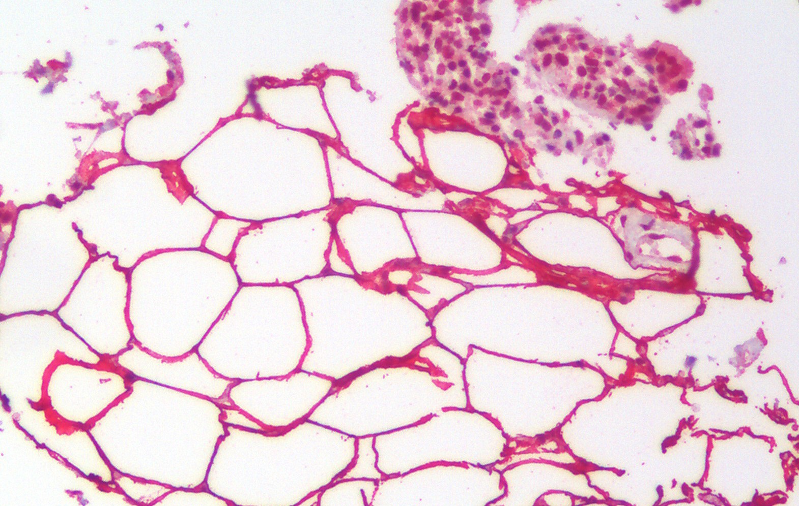
ApplicationsImmunoHistoChemistry
ReactivityHuman, Mouse
TargetGLI1
- SizePrice

Product group Antibodies
Anti-GLI1 AntibodyAMAB91773
ApplicationsImmunoCytoChemistry, ImmunoHistoChemistry
ReactivityHuman, Mouse
TargetGLI1
- SizePrice
Product group Antibodies

GLI / GLI1 AntibodyLS-C834930
ApplicationsWestern Blot, ELISA, ImmunoHistoChemistry
ReactivityHuman, Mouse
TargetGLI1
- SizePrice

Product group Antibodies
Anti-GLI1 Antibody Picoband(r)A00527-3-CARRIER-FREE
ApplicationsWestern Blot, ELISA
ReactivityHuman, Mouse, Rat
TargetGLI1
- SizePrice
Product group Antibodies
References
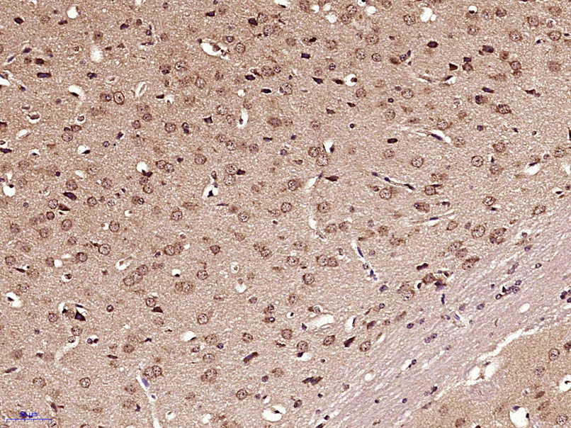
GLI1 Polyclonal AntibodyBS-1206R
ApplicationsFlow Cytometry, ImmunoFluorescence, Western Blot, ELISA, ImmunoCytoChemistry, ImmunoHistoChemistry, ImmunoHistoChemistry Frozen, ImmunoHistoChemistry Paraffin
ReactivityBovine, Canine, Equine, Human, Mouse, Rabbit, Rat
TargetGLI1
- SizePrice
Product group Antibodies
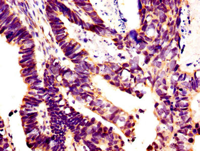
GLI1 AntibodyCSB-PA12989A0RB
ApplicationsImmunoFluorescence, ELISA, ImmunoHistoChemistry
ReactivityHuman
TargetGLI1
- SizePrice