


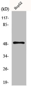
Figure 1. Western blot analysis of GLUT4 using anti-GLUT4 antibody (PB9109). Electrophoresis was performed on a 5-20% SDS-PAGE gel at 70V (Stacking gel) / 90V (Resolving gel) for 2-3 hours. The sample well of each lane was loaded with 50ug of sample under reducing conditions. lane 1: rat cardiac muscle tissue lysate, lane 2: rat skeletal muscle tissue lysate. After Electrophoresis, proteins were transferred to a Nitrocellulose membrane at 150mA for 50-90 minutes. Blocked the membrane with 5% Non-fat Milk/ TBS for 1.5 hour at RT. The membrane was incubated with rabbit anti-GLUT4 antigen affinity purified polyclonal antibody (Catalog # PB9109) at 0.5 microg/mL overnight at 4°C, then washed with TBS-0.1%Tween 3 times with 5 minutes each and probed with a goat anti-rabbit IgG-HRP secondary antibody at a dilution of 1:10000 for 1.5 hour at RT. The signal is developed using an Enhanced Chemiluminescent detection (ECL) kit (Catalog # EK1002) with Tanon 5200 system. A specific band was detected for GLUT4 at approximately 55KD. The expected band size for GLUT4 is at 55KD.
. GLUT4 was detected in frozen section of rat cardiac muscle tissues. The tissue section was blocked with 10% goat serum. The tissue section was then incubated with 1microg/ml rabbit anti-GLUT4 Antibody (PB9109) overnight at 4°C. Biotinylated goat anti-rabbit IgG was used as secondary antibody and incubated for 30 minutes at 37°C. The tissue section was developed using Strepavidin-Biotin-Complex (SABC)(Catalog # SA1022) with DAB as the chromogen.")
. GLUT4 was detected in frozen section of mouse cardiac muscle tissues. The tissue section was blocked with 10% goat serum. The tissue section was then incubated with 1microg/ml rabbit anti-GLUT4 Antibody (PB9109) overnight at 4°C. Biotinylated goat anti-rabbit IgG was used as secondary antibody and incubated for 30 minutes at 37°C. The tissue section was developed using Strepavidin-Biotin-Complex (SABC)(Catalog # SA1022) with DAB as the chromogen.")
. GLUT4 was detected in paraffin-embedded section of human intestinal cancer tissues. Heat mediated antigen retrieval was performed in citrate buffer (pH6, epitope retrieval solution) for 20 mins. The tissue section was blocked with 10% goat serum. The tissue section was then incubated with 1microg/ml rabbit anti-GLUT4 Antibody (PB9109) overnight at 4°C. Biotinylated goat anti-rabbit IgG was used as secondary antibody and incubated for 30 minutes at 37°C. The tissue section was developed using Strepavidin-Biotin-Complex (SABC)(Catalog # SA1022) with DAB as the chromogen.")
. GLUT4 was detected in paraffin-embedded section of rat cardiac muscle tissues. Heat mediated antigen retrieval was performed in citrate buffer (pH6, epitope retrieval solution) for 20 mins. The tissue section was blocked with 10% goat serum. The tissue section was then incubated with 1microg/ml rabbit anti-GLUT4 Antibody (PB9109) overnight at 4°C. Biotinylated goat anti-rabbit IgG was used as secondary antibody and incubated for 30 minutes at 37°C. The tissue section was developed using Strepavidin-Biotin-Complex (SABC)(Catalog # SA1022) with DAB as the chromogen.")
. GLUT4 was detected in paraffin-embedded section of mouse skeletal muscle tissues. Heat mediated antigen retrieval was performed in citrate buffer (pH6, epitope retrieval solution) for 20 mins. The tissue section was blocked with 10% goat serum. The tissue section was then incubated with 1microg/ml rabbit anti-GLUT4 Antibody (PB9109) overnight at 4°C. Biotinylated goat anti-rabbit IgG was used as secondary antibody and incubated for 30 minutes at 37°C. The tissue section was developed using Strepavidin-Biotin-Complex (SABC)(Catalog # SA1022) with DAB as the chromogen.")
Figure 1. Western blot analysis of GLUT4 using anti-GLUT4 antibody (PB9109). Electrophoresis was performed on a 5-20% SDS-PAGE gel at 70V (Stacking gel) / 90V (Resolving gel) for 2-3 hours. The sample well of each lane was loaded with 50ug of sample under reducing conditions. lane 1: rat cardiac muscle tissue lysate, lane 2: rat skeletal muscle tissue lysate. After Electrophoresis, proteins were transferred to a Nitrocellulose membrane at 150mA for 50-90 minutes. Blocked the membrane with 5% Non-fat Milk/ TBS for 1.5 hour at RT. The membrane was incubated with rabbit anti-GLUT4 antigen affinity purified polyclonal antibody (Catalog # PB9109) at 0.5 microg/mL overnight at 4°C, then washed with TBS-0.1%Tween 3 times with 5 minutes each and probed with a goat anti-rabbit IgG-HRP secondary antibody at a dilution of 1:10000 for 1.5 hour at RT. The signal is developed using an Enhanced Chemiluminescent detection (ECL) kit (Catalog # EK1002) with Tanon 5200 system. A specific band was detected for GLUT4 at approximately 55KD. The expected band size for GLUT4 is at 55KD.
Anti-GLUT4 Picoband Antibody
PB9109
ApplicationsWestern Blot, ImmunoHistoChemistry
Product group Antibodies
ReactivityHuman, Mouse, Rat
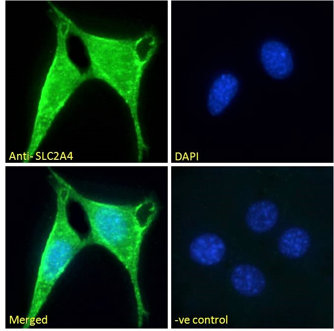
TargetSLC2A4
Overview
- SupplierBoster Bio
- Product NameAnti-GLUT4 Picoband Antibody
- Delivery Days Customer9
- Application Supplier NoteWB: The detection limit for GLUT4 is approximately 0.25ng/lane under reducing conditions. Tested Species: In-house tested species with positive results. Predicted Species: Species predicted to be fit for the product based on sequence similarities. By Heat: Boiling the paraffin sections in 10mM citrate buffer, pH6.0, for 20mins is required for the staining of formalin/paraffin sections. Other applications have not been tested. Optimal dilutions should be determined by end users.
- ApplicationsWestern Blot, ImmunoHistoChemistry
- Applications SupplierIHP, IHF, WB, IHC
- CertificationResearch Use Only
- ClonalityPolyclonal
- Concentration500 ug/ml
- Gene ID6517
- Target nameSLC2A4
- Target descriptionsolute carrier family 2 member 4
- Target synonymsGLUT4, solute carrier family 2, facilitated glucose transporter member 4, GLUT-4, glucose transporter type 4, insulin-responsive, insulin-responsive glucose transporter type 4, solute carrier family 2 (facilitated glucose transporter), member 4
- HostRabbit
- IsotypeIgG
- Protein IDP14672
- Protein NameSolute carrier family 2, facilitated glucose transporter member 4
- Scientific DescriptionBoster Bio Anti-Glucose Transporter GLUT4/SLC2A4 Antibody Picoband® catalog # PB9109. Tested in IHC, WB applications. This antibody reacts with Human, Mouse, Rat. The brand Picoband indicates this is a premium antibody that guarantees superior quality, high affinity, and strong signals with minimal background in Western blot applications. Only our best-performing antibodies are designated as Picoband, ensuring unmatched performance.
- ReactivityHuman, Mouse, Rat
- Reactivity SupplierHuman, Mouse, Rat
- Storage Instruction-20°C,2°C to 8°C
- UNSPSC12352203
References
- Xu L, Zhang C, Bao J, et al. Alpha-lipoic Acid Prevents Bone Loss in Type 2 Diabetes and Postmenopausal Osteoporosis Coexisting Conditions by Modulating the YAP/Glut4 Pathway. Cell Biochem Biophys. 2024,82(2):669-685. doi: 10.1007/s12013-024-01216-wRead this paper
- Zhou J, Wu T, Li C, et al. Alfuzosin ameliorates diabetes by boosting PGK1 activity in diabetic mice. Life Sci. 2023,317:121491. doi: 10.1016/j.lfs.2023.121491Read this paper
- Han X, Yang Y, Liu S, et al. Aerobic exercise ameliorates insulin resistance in C57BL/6 J mice via activating Sestrin3. Biochim Biophys Acta Mol Basis Dis. 2023,1869(1):166568. doi: 10.1016/j.bbadis.2022.166568Read this paper
- Chen X, Zhao H, Meng F, et al. Ameliorated effects of a lipopeptide surfactin on insulin resistance in vitro and in vivo. Food Sci Nutr. 2022,10(7):2455-2469. doi: 10.1002/fsn3.2852Read this paper
- Zhang X, Che L, Shan J, et al. The effects of Formoterol in preventing adipogenesis and obesity are mediated by PPARγ/C/EBPα axis and AMPK/PGC-1α pathway. Biosci Biotechnol Biochem. 2022,:pii: zbac103. doi: 10.1093/bbb/zbac103.Read this paper
- Zhang DS, Liang GY, Liu DX, et al. Role of Phosphorylated AMP-Activated Protein Kinase (AMPK) in Myocardial Insulin Resistance After Myocardial Ischemia-Reperfusion During Cardiopulmonary Bypass Surgery in Dogs. Med Sci Monit. 2019,25:4149-4158. doi: 10.12659/MSM.916517Read this paper
- Costa TC, Moura FH, Souza RO, et al. Effect of maternal feed restriction in dairy goats at different stages of gestation on skeletal muscle development and energy metabolism of kids at the time of births. Anim Reprod Sci. 2019,206:46-59. doi: 10.1016/j.anireprosci.2019.05.006Read this paper
- Mo Z, Li L, Yu H, et al. Coumarins ameliorate diabetogenic action of dexamethasone via Akt activation and AMPK signaling in skeletal muscle. J Pharmacol Sci. 2019,139(3):151-157. doi: 10.1016/j.jphs.2019.01.001Read this paper
- Dai Y, Wang Z, Quan M, et al. Asiatic acid protests against myocardial ischemia/reperfusion injury via modulation of glycometabolism in rat cardiomyocyte. Drug Des Devel Ther. 2018,12:3573-3582. doi: 10.2147/DDDT.S175116Read this paper
- Liu Y, Liu C, Lu ML, et al. Vibration exercise decreases insulin resistance and modulates the insulin signaling pathway in a type 2 diabetic rat model. Int J Clin Exp Med. 2015,8(8):13136-44.Read this paper
Datasheet
MSDS
Related products
Product group Antibodies
ApplicationsImmunoFluorescence, ELISA
ReactivityHuman, Mouse
- SizePrice
Product group Antibodies
SLC2A4 AntibodyCSB-PA002729
ApplicationsWestern Blot, ELISA, ImmunoHistoChemistry
ReactivityHuman, Mouse, Rat
TargetSLC2A4
- SizePrice
Product group Antibodies

SLC2A4 / GLUT-4 AntibodyLS-C402350
ApplicationsELISA, ImmunoHistoChemistry
ReactivityHuman, Mouse, Rat
TargetSLC2A4
- SizePrice
Product group Antibodies
TargetSLC2A4
- SizePrice
Product group Antibodies
Anti-Glucose Transporter GLUT4/SLC2A4 Antibody Picoband(r)PB9109-CARRIER-FREE
ApplicationsWestern Blot, ImmunoHistoChemistry
ReactivityHuman, Mouse, Rat
TargetSLC2A4
- SizePrice
Product group Antibodies
References
GLUT4 Polyclonal AntibodyBS-0384R
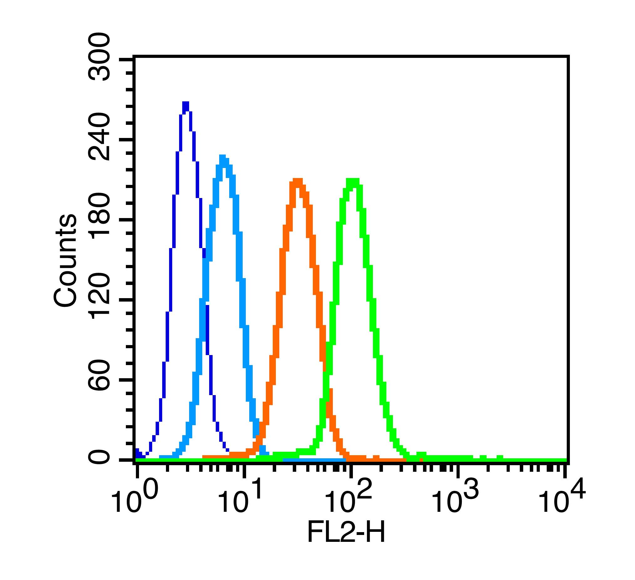
ApplicationsFlow Cytometry, ImmunoFluorescence, Western Blot, ELISA, ImmunoCytoChemistry, ImmunoHistoChemistry, ImmunoHistoChemistry Frozen, ImmunoHistoChemistry Paraffin
ReactivityBovine, Canine, Human, Mouse, Porcine, Rabbit, Rat, Sheep
TargetSLC2A4
- SizePrice
Product group Antibodies

Goat anti-SLC2A4 AntibodyEB10147
ApplicationsImmunoFluorescence, ELISA
ReactivityBovine, Canine, Human, Mouse, Porcine, Rabbit, Rat
TargetSLC2A4
- SizePrice
Product group Antibodies
Anti-SLC2A4 Antibody144-07637
ApplicationsWestern Blot, ImmunoHistoChemistry
ReactivityHuman, Mouse, Rat
TargetSLC2A4
- SizePrice