


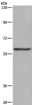
Figure 1. Western blot analysis of Glutaminase/GLS using anti-Glutaminase/GLS antibody (A01272-2). Electrophoresis was performed on a 5-20% SDS-PAGE gel at 70V (Stacking gel) / 90V (Resolving gel) for 2-3 hours. The sample well of each lane was loaded with 50ug of sample under reducing conditions. Lane 1: human placenta tissue lysates, Lane 2: human U87 whole cell lysates, Lane 3: human Hela whole cell lysates, Lane 4: monkey kidney tissue lysates, Lane 5: rat brain tissue lysates, Lane 6: rat kidney tissue lysates, Lane 7: rat heart tissue lysates. Lane 8: rat skeletal muscle tissue lysates. Lane 9: mouse brain tissue lysates. Lane 10: mouse kidney tissue lysates. Lane 11: mouse heart tissue lysates. Lane 12: mouse skeletal muscle tissue lysates. Lane 13: mouse Neuro-2a whole cell lysates. After Electrophoresis, proteins were transferred to a Nitrocellulose membrane at 150mA for 50-90 minutes. Blocked the membrane with 5% Non-fat Milk/ TBS for 1.5 hour at RT. The membrane was incubated with rabbit anti-Glutaminase/GLS antigen affinity purified polyclonal antibody (Catalog # A01272-2) at 0.25 microg/mL overnight at 4°C, then washed with TBS-0.1%Tween 3 times with 5 minutes each and probed with a goat anti-rabbit IgG-HRP secondary antibody at a dilution of 1:10000 for 1.5 hour at RT. The signal is developed using an Enhanced Chemiluminescent detection (ECL) kit (Catalog # EK1002) with Tanon 5200 system. A specific band was detected for Glutaminase/GLS at approximately 65-73KD. The expected band size for Glutaminase/GLS is at 73KD.
. Glutaminase/GLS was detected in paraffin-embedded section of human gastric cancer tissue. Heat mediated antigen retrieval was performed in EDTA buffer (pH8.0, epitope retrieval solution). The tissue section was blocked with 10% goat serum. The tissue section was then incubated with 1microg/ml rabbit anti-Glutaminase/GLS Antibody (A01272-2) overnight at 4°C. Biotinylated goat anti-rabbit IgG was used as secondary antibody and incubated for 30 minutes at 37°C. The tissue section was developed using Strepavidin-Biotin-Complex (SABC) (Catalog # SA1022) with DAB as the chromogen.")
. Glutaminase/GLS was detected in paraffin-embedded section of rat brain tissue. Heat mediated antigen retrieval was performed in EDTA buffer (pH8.0, epitope retrieval solution). The tissue section was blocked with 10% goat serum. The tissue section was then incubated with 1microg/ml rabbit anti-Glutaminase/GLS Antibody (A01272-2) overnight at 4°C. Biotinylated goat anti-rabbit IgG was used as secondary antibody and incubated for 30 minutes at 37°C. The tissue section was developed using Strepavidin-Biotin-Complex (SABC) (Catalog # SA1022) with DAB as the chromogen.")
. Glutaminase/GLS was detected in immunocytochemical section of U20S cells. Enzyme antigen retrieval was performed using IHC enzyme antigen retrieval reagent (AR0022) for 15 mins. The cells were blocked with 10% goat serum. And then incubated with 4microg/mL rabbit anti-Glutaminase/GLS Antibody (A01272-2) overnight at 4°C. DyLight®488 Conjugated Goat Anti-Rabbit IgG (BA1127) was used as secondary antibody at 1:100 dilution and incubated for 30 minutes at 37°C. The section was counterstained with DAPI. Visualize using a fluorescence microscope and filter sets appropriate for the label used.")
. Overlay histogram showing 293T cells stained with A01272-2 (Blue line). To facilitate intracellular staining, cells were fixed with 4% paraformaldehyde and permeabilized with permeabilization buffer. The cells were blocked with 10% normal goat serum. And then incubated with rabbit anti-Glutaminase/GLS Antibody (A01272-2,1microg/1x106 cells) for 30 min at 20°C. DyLight®488 conjugated goat anti-rabbit IgG (BA1127, 5-10microg/1x106 cells) was used as secondary antibody for 30 minutes at 20°C. Isotype control antibody (Green line) was rabbit IgG (1microg/1x106) used under the same conditions. Unlabelled sample without incubation with primary antibody and secondary antibody (Red line) was used as a blank control.")
. Overlay histogram showing SiHa cells stained with A01272-2 (Blue line). To facilitate intracellular staining, cells were fixed with 4% paraformaldehyde and permeabilized with permeabilization buffer. The cells were blocked with 10% normal goat serum. And then incubated with rabbit anti-Glutaminase/GLS Antibody (A01272-2,1microg/1x106 cells) for 30 min at 20°C. DyLight®488 conjugated goat anti-rabbit IgG (BA1127, 5-10microg/1x106 cells) was used as secondary antibody for 30 minutes at 20°C. Isotype control antibody (Green line) was rabbit IgG (1microg/1x106) used under the same conditions. Unlabelled sample without incubation with primary antibody and secondary antibody (Red line) was used as a blank control.")
. GLS was detected in a paraffin-embedded section of human liver cancer tissue. Heat mediated antigen retrieval was performed in EDTA buffer (pH 8.0, epitope retrieval solution). The tissue section was blocked with 10% goat serum. The tissue section was then incubated with 5 microg/ml rabbit anti-GLS Antibody (A01272-2) overnight at 4°C. HRP Conjugated Goat Anti-rabbit IgG was used as secondary antibody and incubated for 30 minutes at 37°C. The tissue section was developed using HRP Conjugated Rabbit IgG Super Vision Assay Kit (Catalog # SV0002) with DAB as the chromogen.")
. GLS was detected in a paraffin-embedded section of mouse brain tissue. Heat mediated antigen retrieval was performed in EDTA buffer (pH 8.0, epitope retrieval solution). The tissue section was blocked with 10% goat serum. The tissue section was then incubated with 5 microg/ml rabbit anti-GLS Antibody (A01272-2) overnight at 4°C. HRP Conjugated Goat Anti-rabbit IgG was used as secondary antibody and incubated for 30 minutes at 37°C. The tissue section was developed using HRP Conjugated Rabbit IgG Super Vision Assay Kit (Catalog # SV0002) with DAB as the chromogen.")
. GLS was detected in a paraffin-embedded section of mouse kidney tissue. Heat mediated antigen retrieval was performed in EDTA buffer (pH 8.0, epitope retrieval solution). The tissue section was blocked with 10% goat serum. The tissue section was then incubated with 5 microg/ml rabbit anti-GLS Antibody (A01272-2) overnight at 4°C. HRP Conjugated Goat Anti-rabbit IgG was used as secondary antibody and incubated for 30 minutes at 37°C. The tissue section was developed using HRP Conjugated Rabbit IgG Super Vision Assay Kit (Catalog # SV0002) with DAB as the chromogen.")
. GLS was detected in a paraffin-embedded section of human endometrial cancer tissue. Heat mediated antigen retrieval was performed in EDTA buffer (pH 8.0, epitope retrieval solution). The tissue section was blocked with 10% goat serum. The tissue section was then incubated with 5 microg/mL rabbit anti-GLS Antibody (A01272-2) overnight at 4°C. DyLight®594 Conjugated Goat Anti-Rabbit IgG (BA1142) was used as secondary antibody at 1:100 dilution and incubated for 30 minutes at 37°C. The section was counterstained with DAPI. Visualize using a fluorescence microscope and filter sets appropriate for the label used.")
Figure 1. Western blot analysis of Glutaminase/GLS using anti-Glutaminase/GLS antibody (A01272-2). Electrophoresis was performed on a 5-20% SDS-PAGE gel at 70V (Stacking gel) / 90V (Resolving gel) for 2-3 hours. The sample well of each lane was loaded with 50ug of sample under reducing conditions. Lane 1: human placenta tissue lysates, Lane 2: human U87 whole cell lysates, Lane 3: human Hela whole cell lysates, Lane 4: monkey kidney tissue lysates, Lane 5: rat brain tissue lysates, Lane 6: rat kidney tissue lysates, Lane 7: rat heart tissue lysates. Lane 8: rat skeletal muscle tissue lysates. Lane 9: mouse brain tissue lysates. Lane 10: mouse kidney tissue lysates. Lane 11: mouse heart tissue lysates. Lane 12: mouse skeletal muscle tissue lysates. Lane 13: mouse Neuro-2a whole cell lysates. After Electrophoresis, proteins were transferred to a Nitrocellulose membrane at 150mA for 50-90 minutes. Blocked the membrane with 5% Non-fat Milk/ TBS for 1.5 hour at RT. The membrane was incubated with rabbit anti-Glutaminase/GLS antigen affinity purified polyclonal antibody (Catalog # A01272-2) at 0.25 microg/mL overnight at 4°C, then washed with TBS-0.1%Tween 3 times with 5 minutes each and probed with a goat anti-rabbit IgG-HRP secondary antibody at a dilution of 1:10000 for 1.5 hour at RT. The signal is developed using an Enhanced Chemiluminescent detection (ECL) kit (Catalog # EK1002) with Tanon 5200 system. A specific band was detected for Glutaminase/GLS at approximately 65-73KD. The expected band size for Glutaminase/GLS is at 73KD.
Anti-Glutaminase/GLS Antibody Picoband(r)
A01272-2-CARRIER-FREE
ApplicationsFlow Cytometry, ImmunoFluorescence, Western Blot, ELISA, ImmunoCytoChemistry, ImmunoHistoChemistry
Product group Antibodies
ReactivityHuman, Monkey, Mouse, Rat
TargetGLS
Overview
- SupplierBoster Bio
- Product NameAnti-Glutaminase/GLS Antibody Picoband(r)
- Delivery Days Customer9
- ApplicationsFlow Cytometry, ImmunoFluorescence, Western Blot, ELISA, ImmunoCytoChemistry, ImmunoHistoChemistry
- CertificationResearch Use Only
- ClonalityPolyclonal
- Concentration500 ug/ml
- Gene ID2744
- Target nameGLS
- Target descriptionglutaminase
- Target synonymsAAD20, CASGID, DEE71, EIEE71, GAC, GAM, GDPAG, GLS1, KGA, glutaminase kidney isoform, mitochondrial, K-glutaminase, L-glutamine amidohydrolase, glutaminase C, glutaminase, phosphate-activated
- HostRabbit
- IsotypeIgG
- Protein IDO94925
- Protein NameGlutaminase kidney isoform, mitochondrial
- Scientific DescriptionBoster Bio Anti-Glutaminase/GLS Antibody Picoband® catalog # A01272-2. Tested in ELISA, Flow Cytometry, IF, IHC, ICC, WB applications. This antibody reacts with Human, Monkey, Mouse, Rat. The brand Picoband indicates this is a premium antibody that guarantees superior quality, high affinity, and strong signals with minimal background in Western blot applications. Only our best-performing antibodies are designated as Picoband, ensuring unmatched performance.
- ReactivityHuman, Monkey, Mouse, Rat
- Storage Instruction-20°C,2°C to 8°C
- UNSPSC12352203
Related products
Product group Antibodies
Anti-GLS AntibodyA39155
ApplicationsWestern Blot, ImmunoHistoChemistry
ReactivityHuman
- SizePrice
Product group Antibodies
Anti-GLS Antibody144-03885
ApplicationsWestern Blot, ImmunoHistoChemistry
ReactivityHuman, Mouse, Rat
TargetGLS
- SizePrice

Product group Antibodies

GLS / Glutaminase AntibodyLS-C831065
ApplicationsWestern Blot, ELISA, ImmunoHistoChemistry
ReactivityHuman, Mouse, Rat
TargetGLS
- SizePrice
Product group Antibodies
GLS1 Polyclonal AntibodyBS-10341R
ApplicationsImmunoFluorescence, Western Blot, ELISA, ImmunoHistoChemistry, ImmunoHistoChemistry Frozen, ImmunoHistoChemistry Paraffin
ReactivityHuman, Mouse, Rat
TargetGLS
- SizePrice
Product group Antibodies
GLS AntibodyCSB-PA009528DA01HU
ApplicationsWestern Blot, ELISA, ImmunoHistoChemistry
ReactivityHuman, Mouse
TargetGLS
- SizePrice
Product group Antibodies
GLS Polyclonal AntibodyCAC13047
ApplicationsWestern Blot, ELISA, ImmunoHistoChemistry
ReactivityMouse
TargetGLS
- SizePrice
![Glutaminase antibody [C1C3] detects Glutaminase protein by Western blot analysis. A. 20 μg Raw264 whole cell lysate/extract Glutaminase antibody [C1C3] (GTX114982) dilution: 1:1000](https://www.genetex.com/upload/website/prouct_img/normal/GTX114982/GTX114982_40240_CT_WB_M_w_23060519_282.webp)
Product group Antibodies
Glutaminase antibody [C1C3]GTX114982
ApplicationsWestern Blot
ReactivityHuman, Mouse
TargetGLS
- SizePrice

Product group Antibodies
Anti-GLS-25ulHPA036223
ApplicationsWestern Blot, ImmunoCytoChemistry, ImmunoHistoChemistry
ReactivityHuman
- SizePrice