


Figure 2. IHC analysis of GNB1 using anti-GNB1 antibody (PB10067). GNB1 was detected in paraffin-embedded section of mouse kidney tissue. Heat mediated antigen retrieval was performed in citrate buffer (pH6, epitope retrieval solution) for 20 mins. The tissue section was blocked with 10% goat serum. The tissue section was then incubated with 1microg/ml rabbit anti-GNB1 Antibody (PB10067) overnight at 4°C. Biotinylated goat anti-rabbit IgG was used as secondary antibody and incubated for 30 minutes at 37°C. The tissue section was developed using Strepavidin-Biotin-Complex (SABC)(Catalog # SA1022) with DAB as the chromogen.
. GNB1 was detected in paraffin-embedded section of rat kidney tissue. Heat mediated antigen retrieval was performed in citrate buffer (pH6, epitope retrieval solution) for 20 mins. The tissue section was blocked with 10% goat serum. The tissue section was then incubated with 1microg/ml rabbit anti-GNB1 Antibody (PB10067) overnight at 4°C. Biotinylated goat anti-rabbit IgG was used as secondary antibody and incubated for 30 minutes at 37°C. The tissue section was developed using Strepavidin-Biotin-Complex (SABC)(Catalog # SA1022) with DAB as the chromogen.")
. GNB1 was detected in paraffin-embedded section of human liver cancer tissue. Heat mediated antigen retrieval was performed in citrate buffer (pH6, epitope retrieval solution) for 20 mins. The tissue section was blocked with 10% goat serum. The tissue section was then incubated with 1microg/ml rabbit anti-GNB1 Antibody (PB10067) overnight at 4°C. Biotinylated goat anti-rabbit IgG was used as secondary antibody and incubated for 30 minutes at 37°C. The tissue section was developed using Strepavidin-Biotin-Complex (SABC)(Catalog # SA1022) with DAB as the chromogen.")
. Overlay histogram showing U937 cells stained with PB10067 (Blue line). To facilitate intracellular staining, cells were fixed with 4% paraformaldehyde and permeabilized with permeabilization buffer. The cells were blocked with 10% normal goat serum. And then incubated with rabbit anti-GNB1 Antibody (PB10067,1microg/1x106 cells) for 30 min at 20°C. DyLight®488 conjugated goat anti-rabbit IgG (BA1127, 5-10microg/1x106 cells) was used as secondary antibody for 30 minutes at 20°C. Isotype control antibody (Green line) was rabbit IgG (1microg/1x106) used under the same conditions. Unlabelled sample without incubation with primary antibody and secondary antibody (Red line) was used as a blank control.")
. GNB1 was detected in immunocytochemical section of A549 cell. Enzyme antigen retrieval was performed using IHC enzyme antigen retrieval reagent (AR0022) for 15 mins. The cells were blocked with 10% goat serum. And then incubated with 2microg/mL rabbit anti-GNB1 Antibody (PB10067) overnight at 4°C. DyLight®488 Conjugated Goat Anti-Rabbit IgG (BA1127) was used as secondary antibody at 1:100 dilution and incubated for 30 minutes at 37°C. The section was counterstained with DAPI. Visualize using a fluorescence microscope and filter sets appropriate for the label used.")
. GNB1 was detected in frozen section of human placenta tissue. The tissue section was blocked with 10% goat serum. The tissue section was then incubated with 1microg/ml rabbit anti-GNB1 Antibody (PB10067) overnight at 4°C. Biotinylated goat anti-rabbit IgG was used as secondary antibody and incubated for 30 minutes at 37°C. The tissue section was developed using Strepavidin-Biotin-Complex (SABC)(Catalog # SA1022) with DAB as the chromogen.")
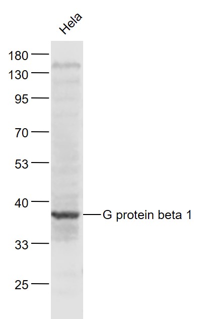
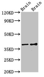
. Electrophoresis was performed on a 5-20% SDS-PAGE gel at 70V (Stacking gel) / 90V (Resolving gel) for 2-3 hours. The sample well of each lane was loaded with 30 ug of sample under reducing conditions. Lane 1: human Hela whole cell lysates, Lane 2: human MCF-7 whole cell lysates, Lane 3: human SiHa whole cell lysates, Lane 4: rat brain tissue lysates, Lane 5: rat liver tissue lysates, Lane 6: mouse brain tissue lysates, Lane 7: mouse liver tissue lysates. After electrophoresis, proteins were transferred to a nitrocellulose membrane at 150 mA for 50-90 minutes. Blocked the membrane with 5% non-fat milk/TBS for 1.5 hour at RT. The membrane was incubated with rabbit anti-GNB1 antigen affinity purified polyclonal antibody (Catalog # PB10067) at 0.5 microg/mL overnight at 4°C, then washed with TBS-0.1%Tween 3 times with 5 minutes each and probed with a goat anti-rabbit IgG-DyLight 647 Conjugated secondary antibody at a dilution of 1:5000 for 1.5 hour at RT. A specific band was detected for GNB1 at approximately 37 kDa. The expected band size for GNB1 is at 37 kDa.")
Figure 2. IHC analysis of GNB1 using anti-GNB1 antibody (PB10067). GNB1 was detected in paraffin-embedded section of mouse kidney tissue. Heat mediated antigen retrieval was performed in citrate buffer (pH6, epitope retrieval solution) for 20 mins. The tissue section was blocked with 10% goat serum. The tissue section was then incubated with 1microg/ml rabbit anti-GNB1 Antibody (PB10067) overnight at 4°C. Biotinylated goat anti-rabbit IgG was used as secondary antibody and incubated for 30 minutes at 37°C. The tissue section was developed using Strepavidin-Biotin-Complex (SABC)(Catalog # SA1022) with DAB as the chromogen.
Anti-GNB1 Antibody Picoband(r)
PB10067-CARRIER-FREE
ApplicationsFlow Cytometry, ImmunoFluorescence, Western Blot, ImmunoCytoChemistry, ImmunoHistoChemistry, ImmunoHistoChemistry Frozen
Product group Antibodies
ReactivityBovine, Canine, Equine, Hamster, Human, Monkey, Mouse, Rabbit, Rat, Zebra Fish
TargetGNB1
Overview
- SupplierBoster Bio
- Product NameAnti-GNB1 Antibody Picoband(r)
- Delivery Days Customer9
- Application Supplier NoteTested Species: In-house tested species with positive results. By Heat: Boiling the paraffin sections in 10mM citrate buffer, pH6.0, for 20mins is required for the staining of formalin/paraffin sections. Other applications have not been tested. Optimal dilutions should be determined by end users.
- ApplicationsFlow Cytometry, ImmunoFluorescence, Western Blot, ImmunoCytoChemistry, ImmunoHistoChemistry, ImmunoHistoChemistry Frozen
- CertificationResearch Use Only
- ClonalityPolyclonal
- Concentration500 ug/ml
- Gene ID2782
- Target nameGNB1
- Target descriptionG protein subunit beta 1
- Target synonymsHG2A, MDS, MRD42, guanine nucleotide-binding protein G(I)/G(S)/G(T) subunit beta-1, beta subunit, signal-transducing proteins GS/GI, guanine nucleotide binding protein (G protein), beta polypeptide 1, heterotrimeric guanine nucleotide-binding protein 2A, testicular tissue protein Li 72, transducin beta chain 1
- HostRabbit
- IsotypeIgG
- Protein IDP62873
- Protein NameGuanine nucleotide-binding protein G(I)/G(S)/G(T) subunit beta-1
- Scientific DescriptionBoster Bio Anti-GNB1 Antibody Picoband® catalog # PB10067. Tested in Flow Cytometry, IF, IHC, IHC-F, ICC, WB applications. This antibody reacts with Human, Mouse, Rat. The brand Picoband indicates this is a premium antibody that guarantees superior quality, high affinity, and strong signals with minimal background in Western blot applications. Only our best-performing antibodies are designated as Picoband, ensuring unmatched performance.
- ReactivityBovine, Canine, Equine, Hamster, Human, Monkey, Mouse, Rabbit, Rat, Zebra Fish
- Storage Instruction-20°C,2°C to 8°C
- UNSPSC12352203
Related products
Product group Antibodies
Anti-GNB1 AntibodyA31216
ApplicationsWestern Blot, ImmunoHistoChemistry
ReactivityHuman, Mouse, Rat
- SizePrice
Product group Antibodies
Anti-GNB1 Antibody144-01867
ApplicationsWestern Blot
ReactivityHuman, Mouse, Rat
TargetGNB1
- SizePrice
Product group Antibodies
ApplicationsImmunoFluorescence, Western Blot, ELISA, ImmunoCytoChemistry, ImmunoHistoChemistry, ImmunoHistoChemistry Frozen, ImmunoHistoChemistry Paraffin
ReactivityBovine, Chicken, Human, Mouse, Porcine, Rat, Sheep
TargetGNB1
- SizePrice
Product group Antibodies
GNB1 AntibodyCSB-PA009602LA01HU
ApplicationsImmunoFluorescence, Western Blot, ELISA
ReactivityHuman, Mouse, Rat
TargetGNB1
- SizePrice
Product group Antibodies
GNB1 Polyclonal AntibodyCAC14840
ApplicationsImmunoFluorescence, Western Blot, ELISA
ReactivityMouse, Rat
TargetGNB1
- SizePrice
Product group Antibodies

GNB1 AntibodyLS-C400755
ApplicationsWestern Blot, ELISA, ImmunoHistoChemistry
ReactivityHuman, Mouse, Rat
TargetGNB1
- SizePrice
Product group Antibodies
GNB1 antibodyGTX114442
ApplicationsImmunoFluorescence, ImmunoPrecipitation, Western Blot, ImmunoCytoChemistry, ImmunoHistoChemistry, ImmunoHistoChemistry Frozen, ImmunoHistoChemistry Paraffin, Other Application
ReactivityHuman, Mouse, Rat, Zebra Fish
TargetGNB1
- SizePrice
Product group Antibodies
ApplicationsImmunoFluorescence, ImmunoPrecipitation, Western Blot, ELISA, ImmunoCytoChemistry, ImmunoHistoChemistry, ImmunoHistoChemistry Paraffin
ReactivityHuman
TargetGNB1
- SizePrice