


Immunohistochemical staining of human liver shows no positivity in hepatocytes as expected.
Immunohistochemical staining of human liver shows no positivity in hepatocytes as expected.
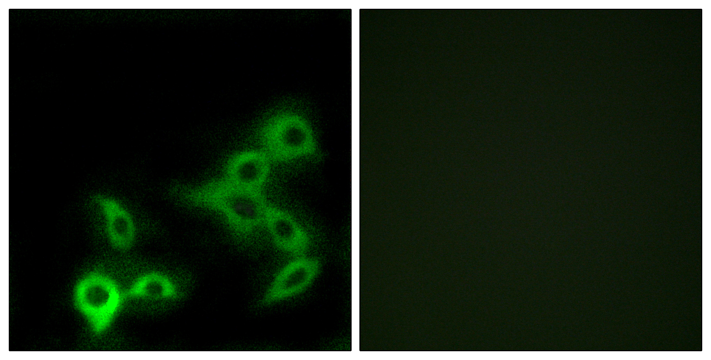
Anti-GPBAR1 Antibody
HPA062890
ApplicationsImmunoHistoChemistry
Product group Antibodies
ReactivityHuman
TargetGPBAR1
Overview
- SupplierAtlas Antibodies
- Product NameAnti-GPBAR1 Antibody
- Delivery Days Customer4
- ApplicationsImmunoHistoChemistry
- CertificationResearch Use Only
- ClonalityPolyclonal
- ConjugateUnconjugated
- Gene ID151306
- Target nameGPBAR1
- Target descriptionG protein-coupled bile acid receptor 1
- Target synonymsBG37, GPCR19, GPR131, M-BAR, TGR5, G-protein coupled bile acid receptor 1, G-protein coupled bile acid receptor BG37, G-protein coupled receptor GPCR19, membrane bile acid receptor, membrane-type receptor for bile acids
- HostRabbit
- IsotypeIgG
- Protein IDQ8TDU6
- Protein NameG-protein coupled bile acid receptor 1
- Scientific DescriptionRecombinant Protein Epitope Signature Tag (PrEST) antigen sequence
- ReactivityHuman
- Storage Instruction-20°C,2°C to 8°C
- UNSPSC41116161
Datasheet
MSDS
Related products
Product group Antibodies

GPBAR AntibodyABX015316
ApplicationsImmunoFluorescence, ELISA, ImmunoCytoChemistry
- SizePrice
Product group Antibodies
Anti-GPBAR AntibodyA101073
ApplicationsImmunoFluorescence, ELISA
ReactivityHuman
- SizePrice

Product group Antibodies
Anti-GPCR TGR5/GPBAR1 Antibody Picoband(r)A01958-1-CARRIER-FREE
ApplicationsWestern Blot
ReactivityMouse, Rat
TargetGPBAR1
- SizePrice
Product group Antibodies
GPBAR1 Polyclonal AntibodyBS-8874R
ApplicationsImmunoFluorescence, Western Blot, ImmunoHistoChemistry, ImmunoHistoChemistry Paraffin
ReactivityHuman
TargetGPBAR1
- SizePrice
Product group Antibodies
GPBAR1 AntibodyCSB-PA007456
ApplicationsImmunoFluorescence, ELISA
ReactivityHuman
TargetGPBAR1
- SizePrice
Product group Antibodies

ApplicationsImmunoFluorescence, ELISA
ReactivityHuman
TargetGPBAR1
- SizePrice