


Immunohistochemical staining of human small intestine shows strong membranous positivity in glandular cells.
Immunohistochemical staining of human small intestine shows strong membranous positivity in glandular cells.
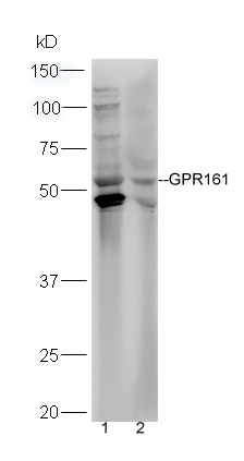
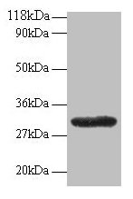
Anti-GPR161 Antibody
HPA015576
ApplicationsImmunoHistoChemistry
Product group Antibodies
ReactivityHuman
TargetGPR161
Overview
- SupplierAtlas Antibodies
- Product NameAnti-GPR161 Antibody
- Delivery Days Customer4
- ApplicationsImmunoHistoChemistry
- CertificationResearch Use Only
- ClonalityPolyclonal
- ConjugateUnconjugated
- Gene ID23432
- Target nameGPR161
- Target descriptionG protein-coupled receptor 161
- Target synonymsRE2, G-protein coupled receptor 161, G-protein coupled receptor RE2
- HostRabbit
- IsotypeIgG
- Protein IDQ8N6U8
- Protein NameG-protein coupled receptor 161
- Scientific DescriptionRecombinant Protein Epitope Signature Tag (PrEST) antigen sequence
- ReactivityHuman
- Storage Instruction-20°C,2°C to 8°C
- UNSPSC41116161
Datasheet
MSDS
Related products

Product group Antibodies
Anti-GPR161 Antibody Picoband(r)A10567-1-CARRIER-FREE
ApplicationsWestern Blot
ReactivityHuman, Mouse, Rat
TargetGPR161
- SizePrice
Product group Antibodies

GPR161 AntibodyLS-C830128
ApplicationsELISA, ImmunoHistoChemistry
ReactivityHuman, Mouse
TargetGPR161
- SizePrice
Product group Antibodies
GPR161 Polyclonal AntibodyBS-15385R
ApplicationsImmunoFluorescence, Western Blot, ELISA, ImmunoCytoChemistry, ImmunoHistoChemistry, ImmunoHistoChemistry Frozen, ImmunoHistoChemistry Paraffin
ReactivityBovine, Canine, Equine, Human, Mouse, Porcine, Rat, Sheep
TargetGPR161
- SizePrice
Product group Antibodies
GPR161 Polyclonal AntibodyCAC14056
ApplicationsImmunoFluorescence, Western Blot, ELISA
TargetGPR161
- SizePrice
Product group Antibodies
GPR161 AntibodyCSB-PA04114A0RB
ApplicationsImmunoFluorescence, Western Blot, ELISA
ReactivityHuman
TargetGPR161
- SizePrice

Product group Antibodies
Anti-GPR161 AntibodyHPA072047
ApplicationsImmunoCytoChemistry
ReactivityHuman
TargetGPR161
- SizePrice