


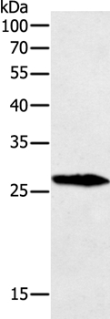
Figure 1. Western blot analysis of GREM1 using anti-GREM1 antibody (PB9626). Electrophoresis was performed on a 5-20% SDS-PAGE gel at 70V (Stacking gel) / 90V (Resolving gel) for 2-3 hours. The sample well of each lane was loaded with 30 ug of sample under reducing conditions. Lane 1: human A549 whole cell lysates, Lane 2: rat testis tissue lysates, Lane 3: mouse testis tissue lysates. After electrophoresis, proteins were transferred to a nitrocellulose membrane at 150 mA for 50-90 minutes. Blocked the membrane with 5% non-fat milk/TBS for 1.5 hour at RT. The membrane was incubated with rabbit anti-GREM1 antigen affinity purified polyclonal antibody (Catalog # PB9626) at 0.5 microg/mL overnight at 4°C, then washed with TBS-0.1%Tween 3 times with 5 minutes each and probed with a goat anti-rabbit IgG-HRP secondary antibody at a dilution of 1:5000 for 1.5 hour at RT. The signal is developed using an Enhanced Chemiluminescent detection (ECL) kit (Catalog # EK1002) with Tanon 5200 system. A specific band was detected for GREM1 at approximately 21 kDa. The expected band size for GREM1 is at 21 kDa.
Figure 1. Western blot analysis of GREM1 using anti-GREM1 antibody (PB9626). Electrophoresis was performed on a 5-20% SDS-PAGE gel at 70V (Stacking gel) / 90V (Resolving gel) for 2-3 hours. The sample well of each lane was loaded with 30 ug of sample under reducing conditions. Lane 1: human A549 whole cell lysates, Lane 2: rat testis tissue lysates, Lane 3: mouse testis tissue lysates. After electrophoresis, proteins were transferred to a nitrocellulose membrane at 150 mA for 50-90 minutes. Blocked the membrane with 5% non-fat milk/TBS for 1.5 hour at RT. The membrane was incubated with rabbit anti-GREM1 antigen affinity purified polyclonal antibody (Catalog # PB9626) at 0.5 microg/mL overnight at 4°C, then washed with TBS-0.1%Tween 3 times with 5 minutes each and probed with a goat anti-rabbit IgG-HRP secondary antibody at a dilution of 1:5000 for 1.5 hour at RT. The signal is developed using an Enhanced Chemiluminescent detection (ECL) kit (Catalog # EK1002) with Tanon 5200 system. A specific band was detected for GREM1 at approximately 21 kDa. The expected band size for GREM1 is at 21 kDa.
Anti-Gremlin 1/GREM1 Antibody Picoband(r)
PB9626-CARRIER-FREE
ApplicationsWestern Blot
Product group Antibodies
ReactivityHamster, Human, Mouse, Rat
TargetGREM1
Overview
- SupplierBoster Bio
- Product NameAnti-Gremlin 1/GREM1 Antibody Picoband(r)
- Delivery Days Customer9
- Application Supplier NoteTested Species: In-house tested species with positive results. Predicted Species: Species predicted to be fit for the product based on sequence similarities. Other applications have not been tested. Optimal dilutions should be determined by end users.
- ApplicationsWestern Blot
- CertificationResearch Use Only
- ClonalityPolyclonal
- Concentration500 ug/ml
- Gene ID26585
- Target nameGREM1
- Target descriptiongremlin 1, DAN family BMP antagonist
- Target synonymsC15DUPq, CKTSF1B1, CRAC1, CRCS4, DAND2, DRM, DUP15q, GREMLIN, HMPS, HMPS1, IHG-2, MPSH, PIG2, gremlin-1, DAN domain family member 2, cell proliferation-inducing gene 2 protein, colorectal adenoma and carcinoma 1, cysteine knot superfamily 1, BMP antagonist 1, down-regulated in Mos-transformed cells protein, gremlin 1, cysteine knot superfamily, homolog, gremlin 1-like protein, hereditary mixed polyposis syndrome, increased in high glucose-2
- HostRabbit
- IsotypeIgG
- Protein IDO60565
- Protein NameGremlin-1
- Scientific DescriptionBoster Bio Anti-Gremlin 1/GREM1 Antibody Picoband® catalog # PB9626. Tested in WB applications. This antibody reacts with Human, Mouse, Rat. The brand Picoband indicates this is a premium antibody that guarantees superior quality, high affinity, and strong signals with minimal background in Western blot applications. Only our best-performing antibodies are designated as Picoband, ensuring unmatched performance.
- ReactivityHamster, Human, Mouse, Rat
- Storage Instruction-20°C,2°C to 8°C
- UNSPSC12352203
Related products
Product group Antibodies
Anti-GREM1 Antibody144-11595
ApplicationsWestern Blot
ReactivityHuman
TargetGREM1
- SizePrice
Product group Antibodies
Anti-GREM1 AntibodyA43148
ApplicationsWestern Blot
ReactivityHuman, Mouse
- SizePrice
Product group Antibodies
Gremlin Polyclonal AntibodyBS-1475R
ApplicationsImmunoFluorescence, Western Blot, ELISA, ImmunoCytoChemistry, ImmunoHistoChemistry, ImmunoHistoChemistry Frozen, ImmunoHistoChemistry Paraffin
ReactivityBovine, Canine, Chicken, Equine, Human, Mouse, Rabbit, Rat
TargetGREM1
- SizePrice
Product group Antibodies
GREM1 AntibodyCSB-PA009892LA01HU
ApplicationsELISA, ImmunoHistoChemistry
ReactivityHuman
TargetGREM1
- SizePrice
Product group Antibodies
ApplicationsImmunoPrecipitation, Western Blot, ImmunoCytoChemistry, ImmunoHistoChemistry
ReactivityMouse, Rat
TargetGREM1
- SizePrice
Product group Antibodies

GREM1 / Gremlin-1 AntibodyLS-C404029
ApplicationsWestern Blot, ELISA
ReactivityHuman, Mouse, Rat
TargetGREM1
- SizePrice
Product group Antibodies
Gremlin 1 antibodyGTX03394
ApplicationsImmunoHistoChemistry, ImmunoHistoChemistry Paraffin
ReactivityHuman
TargetGREM1
- SizePrice