


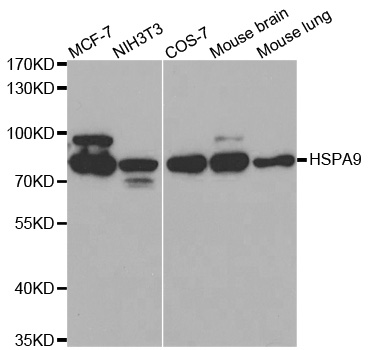
Figure 1. Western blot analysis of Grp75 using anti-Grp75 antibody (M02561-2). Electrophoresis was performed on a 5-20% SDS-PAGE gel at 70V (Stacking gel) / 90V (Resolving gel) for 2-3 hours. The sample well of each lane was loaded with 30 ug of sample under reducing conditions. Lane 1: human Hela whole cell lysates, Lane 2: human HepG2 whole cell lysates, Lane 3: rat brain tissue lysates, Lane 4: rat lung tissue lysates, Lane 5: rat PC-12 whole cell lysates, Lane 6: mouse brain tissue lysates, Lane 7: mouse lung tissue lysates, Lane 8: mouse RAW264.7 whole cell lysates. After electrophoresis, proteins were transferred to a nitrocellulose membrane at 150 mA for 50-90 minutes. Blocked the membrane with 5% non-fat milk/TBS for 1.5 hour at RT. The membrane was incubated with mouse anti-Grp75 antigen affinity purified monoclonal antibody (Catalog # M02561-2) at 0.5 microg/mL overnight at 4°C, then washed with TBS-0.1%Tween 3 times with 5 minutes each and probed with a goat anti-mouse IgG-HRP secondary antibody at a dilution of 1:10000 for 1.5 hour at RT. The signal is developed using an Enhanced Chemiluminescent detection (ECL) kit (Catalog # EK1001) with Tanon 5200 system. A specific band was detected for Grp75 at approximately 74 kDa. The expected band size for Grp75 is at 74 kDa.
. Grp75 was detected in a paraffin-embedded section of human appendiceal adenocarcinoma tissue. Heat mediated antigen retrieval was performed in EDTA buffer (pH 8.0, epitope retrieval solution). The tissue section was blocked with 10% goat serum. The tissue section was then incubated with 2 microg/ml mouse anti-Grp75 Antibody (M02561-2) overnight at 4°C. Biotinylated goat anti-mouse IgG was used as secondary antibody and incubated for 30 minutes at 37°C. The tissue section was developed using Strepavidin-Biotin-Complex (SABC) (Catalog # SA1021) with DAB as the chromogen.")
. Grp75 was detected in a paraffin-embedded section of human gastric cancer tissue. Heat mediated antigen retrieval was performed in EDTA buffer (pH 8.0, epitope retrieval solution). The tissue section was blocked with 10% goat serum. The tissue section was then incubated with 2 microg/ml mouse anti-Grp75 Antibody (M02561-2) overnight at 4°C. Biotinylated goat anti-mouse IgG was used as secondary antibody and incubated for 30 minutes at 37°C. The tissue section was developed using Strepavidin-Biotin-Complex (SABC) (Catalog # SA1021) with DAB as the chromogen.")
. Grp75 was detected in a paraffin-embedded section of human gall bladder adenosquamous carcinoma tissue. Heat mediated antigen retrieval was performed in EDTA buffer (pH 8.0, epitope retrieval solution). The tissue section was blocked with 10% goat serum. The tissue section was then incubated with 2 microg/ml mouse anti-Grp75 Antibody (M02561-2) overnight at 4°C. Biotinylated goat anti-mouse IgG was used as secondary antibody and incubated for 30 minutes at 37°C. The tissue section was developed using Strepavidin-Biotin-Complex (SABC) (Catalog # SA1021) with DAB as the chromogen.")
. Grp75 was detected in a paraffin-embedded section of human lymphadenoma tissue. Heat mediated antigen retrieval was performed in EDTA buffer (pH 8.0, epitope retrieval solution). The tissue section was blocked with 10% goat serum. The tissue section was then incubated with 2 microg/ml mouse anti-Grp75 Antibody (M02561-2) overnight at 4°C. Biotinylated goat anti-mouse IgG was used as secondary antibody and incubated for 30 minutes at 37°C. The tissue section was developed using Strepavidin-Biotin-Complex (SABC) (Catalog # SA1021) with DAB as the chromogen.")
. Grp75 was detected in an immunocytochemical section of CACO-2 cells. Enzyme antigen retrieval was performed using IHC enzyme antigen retrieval reagent (AR0022) for 15 mins. The cells were blocked with 10% goat serum. And then incubated with 5 microg/mL mouse anti-Grp75 Antibody (M02561-2) overnight at 4°C. DyLight®488 Conjugated Goat Anti-Mouse IgG (BA1126) was used as secondary antibody at 1:100 dilution and incubated for 30 minutes at 37°C. The section was counterstained with DAPI. Visualize using a fluorescence microscope and filter sets appropriate for the label used.")
. Overlay histogram showing HepG2 cells stained with M02561-2 (Blue line). To facilitate intracellular staining, cells were fixed with 4% paraformaldehyde and permeabilized with permeabilization buffer. The cells were blocked with 10% normal goat serum. And then incubated with mouse anti-Grp75 Antibody (M02561-2, 1 microg/1x106 cells) for 30 min at 20°C. DyLight®488 conjugated goat anti-mouse IgG (BA1126, 5-10 microg/1x106 cells) was used as secondary antibody for 30 minutes at 20°C. Isotype control antibody (Green line) was mouse IgG (1 microg/1x106) used under the same conditions. Unlabelled sample without incubation with primary antibody and secondary antibody (Red line) was used as a blank control.")
Figure 1. Western blot analysis of Grp75 using anti-Grp75 antibody (M02561-2). Electrophoresis was performed on a 5-20% SDS-PAGE gel at 70V (Stacking gel) / 90V (Resolving gel) for 2-3 hours. The sample well of each lane was loaded with 30 ug of sample under reducing conditions. Lane 1: human Hela whole cell lysates, Lane 2: human HepG2 whole cell lysates, Lane 3: rat brain tissue lysates, Lane 4: rat lung tissue lysates, Lane 5: rat PC-12 whole cell lysates, Lane 6: mouse brain tissue lysates, Lane 7: mouse lung tissue lysates, Lane 8: mouse RAW264.7 whole cell lysates. After electrophoresis, proteins were transferred to a nitrocellulose membrane at 150 mA for 50-90 minutes. Blocked the membrane with 5% non-fat milk/TBS for 1.5 hour at RT. The membrane was incubated with mouse anti-Grp75 antigen affinity purified monoclonal antibody (Catalog # M02561-2) at 0.5 microg/mL overnight at 4°C, then washed with TBS-0.1%Tween 3 times with 5 minutes each and probed with a goat anti-mouse IgG-HRP secondary antibody at a dilution of 1:10000 for 1.5 hour at RT. The signal is developed using an Enhanced Chemiluminescent detection (ECL) kit (Catalog # EK1001) with Tanon 5200 system. A specific band was detected for Grp75 at approximately 74 kDa. The expected band size for Grp75 is at 74 kDa.
Anti-Grp75 Antibody Picoband(r) (monoclonal, 4I9)
M02561-2-IFLUOR647
ApplicationsFlow Cytometry, ImmunoFluorescence, Western Blot, ImmunoCytoChemistry, ImmunoHistoChemistry
Product group Antibodies
ReactivityHuman, Mouse, Rat
TargetHSPA9
Overview
- SupplierBoster Bio
- Product NameAnti-Grp75 Antibody Picoband(r) (monoclonal, 4I9)
- Delivery Days Customer9
- ApplicationsFlow Cytometry, ImmunoFluorescence, Western Blot, ImmunoCytoChemistry, ImmunoHistoChemistry
- CertificationResearch Use Only
- ClonalityMonoclonal
- Clone ID4I9
- Concentration500 ug/ml
- ConjugateOther Conjugate
- Gene ID3313
- Target nameHSPA9
- Target descriptionheat shock protein family A (Hsp70) member 9
- Target synonymsCRP40, CSA, EVPLS, GRP-75, GRP75, HEL-S-124m, HSPA9B, MOT, MOT2, MTHSP75, PBP74, SAAN, SIDBA4, stress-70 protein, mitochondrial, 75 kDa glucose-regulated protein, catecholamine-regulated protein 40, epididymis secretory sperm binding protein Li 124m, heat shock 70kD protein 9B, heat shock 70kDa protein 9 (mortalin), heat shock protein family A member 9, mortalin, perinuclear, mortalin-2, mortalin2, p66-mortalin, peptide-binding protein 74
- HostMouse
- IsotypeIgG1
- Protein IDP38646
- Protein NameStress-70 protein, mitochondrial
- Scientific DescriptionBoster Bio Anti-Grp75 Antibody Picoband® (monoclonal, 4I9) catalog # M02561-2. Tested in Flow Cytometry, IF, IHC, ICC, WB applications. This antibody reacts with Human, Mouse, Rat. The brand Picoband indicates this is a premium antibody that guarantees superior quality, high affinity, and strong signals with minimal background in Western blot applications. Only our best-performing antibodies are designated as Picoband, ensuring unmatched performance.
- ReactivityHuman, Mouse, Rat
- Storage Instruction-20°C,2°C to 8°C
- UNSPSC12352203
Related products
Product group Antibodies
HSPA9 AntibodyPACO06566
ApplicationsImmunoFluorescence, Western Blot, ELISA, ImmunoHistoChemistry
ReactivityHuman, Monkey, Mouse, Rat
TargetHSPA9
- SizePrice
Product group Antibodies
ApplicationsImmunoPrecipitation, Western Blot, ImmunoCytoChemistry, ImmunoHistoChemistry
ReactivityMouse, Porcine
TargetHSPA9
- SizePrice
Product group Antibodies
Anti-HSPA9 Antibody144-00558
ApplicationsImmunoFluorescence, Western Blot, ImmunoHistoChemistry
ReactivityHuman, Mouse, Rat
TargetHSPA9
- SizePrice

Product group Antibodies
Anti-Grp75/HSPA9 Antibody Picoband(r)PB9642-CARRIER-FREE
ApplicationsFlow Cytometry, ImmunoFluorescence, ImmunoPrecipitation, Western Blot, ImmunoCytoChemistry, ImmunoHistoChemistry
ReactivityHuman, Mouse, Rat
TargetHSPA9
- SizePrice
Product group Antibodies
References
Grp75 antibodyGTX104407
ApplicationsImmunoFluorescence, ImmunoPrecipitation, Western Blot, ImmunoCytoChemistry, ImmunoHistoChemistry, ImmunoHistoChemistry Paraffin
ReactivityHuman, Mouse, Porcine, Rat
TargetHSPA9
- SizePrice
Product group Antibodies

HSPA9 AntibodyABX330079
ApplicationsImmunoFluorescence, Western Blot, ELISA, ImmunoCytoChemistry, ImmunoHistoChemistry
- SizePrice
Product group Antibodies
Anti-GRP75 AntibodyA29499
ApplicationsImmunoFluorescence, ImmunoPrecipitation, Western Blot, ImmunoHistoChemistry, Other Application
ReactivityHuman, Mouse, Rat
- SizePrice
Product group Antibodies
HSPA9 AntibodyCSB-PA002982
ApplicationsImmunoFluorescence, Western Blot, ELISA, ImmunoHistoChemistry
ReactivityHuman, Monkey, Mouse, Rat
TargetHSPA9
- SizePrice
Product group Antibodies
GRP75 Recombinant Antibody, AbBy Fluor-350 ConjugatedBSM-61580R-BF350
ApplicationsWestern Blot
ReactivityHuman, Mouse, Rat
TargetHSPA9
- SizePrice