


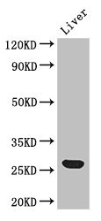
Figure 1. Western blot analysis of GSTA1/A2/A3/A4/A5 using anti-GSTA1/A2/A3/A4/A5 antibody (A01462-1). Electrophoresis was performed on a 5-20% SDS-PAGE gel at 70V (Stacking gel) / 90V (Resolving gel) for 2-3 hours. The sample well of each lane was loaded with 30 ug of sample under reducing conditions. Lane 1: human HepG2 whole cell lysates, Lane 2: human HCCT tissue lysates, Lane 3: human HCCP tissue lysates, Lane 4: human HUH-7 whole cell lysates, Lane 5: rat liver tissue lysates, Lane 6: rat RH35 whole cell lysates, Lane 7: mouse liver tissue lysates. After electrophoresis, proteins were transferred to a nitrocellulose membrane at 150 mA for 50-90 minutes. Blocked the membrane with 5% non-fat milk/TBS for 1.5 hour at RT. The membrane was incubated with rabbit anti-GSTA1/A2/A3/A4/A5 antigen affinity purified polyclonal antibody (Catalog # A01462-1) at 0.5 microg/mL overnight at 4°C, then washed with TBS-0.1%Tween 3 times with 5 minutes each and probed with a goat anti-rabbit IgG-HRP secondary antibody at a dilution of 1:5000 for 1.5 hour at RT. The signal is developed using an Enhanced Chemiluminescent detection (ECL) kit (Catalog # EK1002) with Tanon 5200 system. A specific band was detected for GSTA1/A2/A3/A4/A5 at approximately 26 kDa. The expected band size for GSTA1/A2/A3/A4/A5 is at 26 kDa.
. GSTA1/A2/A3/A4/A5 was detected in paraffin-embedded section of human lung cancer tissues. Heat mediated antigen retrieval was performed in citrate buffer (pH6, epitope retrieval solution) for 20 mins. The tissue section was blocked with 10% goat serum. The tissue section was then incubated with 1microg/ml rabbit anti-GSTA1/A2/A3/A4/A5 Antibody (A01462-1) overnight at 4°C. Biotinylated goat anti-rabbit IgG was used as secondary antibody and incubated for 30 minutes at 37°C. The tissue section was developed using Strepavidin-Biotin-Complex (SABC)(Catalog # SA1022) with DAB as the chromogen.")
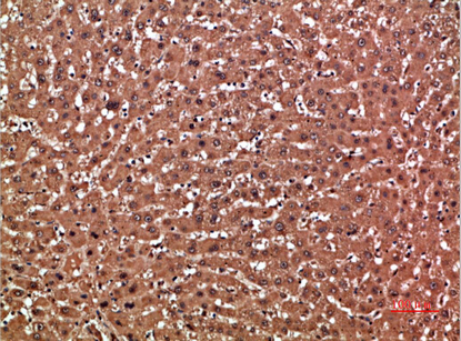
. GSTA1/A2/A3/A4/A5 was detected in paraffin-embedded section of human liver cancer tissues. Heat mediated antigen retrieval was performed in citrate buffer (pH6, epitope retrieval solution) for 20 mins. The tissue section was blocked with 10% goat serum. The tissue section was then incubated with 1microg/ml rabbit anti-GSTA1/A2/A3/A4/A5 Antibody (A01462-1) overnight at 4°C. Biotinylated goat anti-rabbit IgG was used as secondary antibody and incubated for 30 minutes at 37°C. The tissue section was developed using Strepavidin-Biotin-Complex (SABC)(Catalog # SA1022) with DAB as the chromogen.")
. GSTA1/A2/A3/A4/A5 was detected in paraffin-embedded section of human liver cancer tissues. Heat mediated antigen retrieval was performed in citrate buffer (pH6, epitope retrieval solution) for 20 mins. The tissue section was blocked with 10% goat serum. The tissue section was then incubated with 1microg/ml rabbit anti-GSTA1/A2/A3/A4/A5 Antibody (A01462-1) overnight at 4°C. Biotinylated goat anti-rabbit IgG was used as secondary antibody and incubated for 30 minutes at 37°C. The tissue section was developed using Strepavidin-Biotin-Complex (SABC)(Catalog # SA1022) with DAB as the chromogen.")
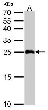
Figure 1. Western blot analysis of GSTA1/A2/A3/A4/A5 using anti-GSTA1/A2/A3/A4/A5 antibody (A01462-1). Electrophoresis was performed on a 5-20% SDS-PAGE gel at 70V (Stacking gel) / 90V (Resolving gel) for 2-3 hours. The sample well of each lane was loaded with 30 ug of sample under reducing conditions. Lane 1: human HepG2 whole cell lysates, Lane 2: human HCCT tissue lysates, Lane 3: human HCCP tissue lysates, Lane 4: human HUH-7 whole cell lysates, Lane 5: rat liver tissue lysates, Lane 6: rat RH35 whole cell lysates, Lane 7: mouse liver tissue lysates. After electrophoresis, proteins were transferred to a nitrocellulose membrane at 150 mA for 50-90 minutes. Blocked the membrane with 5% non-fat milk/TBS for 1.5 hour at RT. The membrane was incubated with rabbit anti-GSTA1/A2/A3/A4/A5 antigen affinity purified polyclonal antibody (Catalog # A01462-1) at 0.5 microg/mL overnight at 4°C, then washed with TBS-0.1%Tween 3 times with 5 minutes each and probed with a goat anti-rabbit IgG-HRP secondary antibody at a dilution of 1:5000 for 1.5 hour at RT. The signal is developed using an Enhanced Chemiluminescent detection (ECL) kit (Catalog # EK1002) with Tanon 5200 system. A specific band was detected for GSTA1/A2/A3/A4/A5 at approximately 26 kDa. The expected band size for GSTA1/A2/A3/A4/A5 is at 26 kDa.
Anti-GSTA1/A2/A3/A4/A5 Antibody Picoband(r)
A01462-1-CARRIER-FREE
ApplicationsWestern Blot, ELISA, ImmunoHistoChemistry
Product group Antibodies
ReactivityHuman, Mouse, Rat
TargetGSTA1
Overview
- SupplierBoster Bio
- Product NameAnti-GSTA1/A2/A3/A4/A5 Antibody Picoband(r)
- Delivery Days Customer9
- ApplicationsWestern Blot, ELISA, ImmunoHistoChemistry
- CertificationResearch Use Only
- ClonalityPolyclonal
- Concentration500 ug/ml
- Gene ID2938
- Target nameGSTA1
- Target descriptionglutathione S-transferase alpha 1
- Target synonymsGST-epsilon, GST2, GSTA1-1, GTH1, glutathione S-transferase A1, 13-hydroperoxyoctadecadienoate peroxidase, GST HA subunit 1, GST class-alpha member 1, S-(hydroxyalkyl)glutathione lyase A1, androst-5-ene-3,17-dione isomerase, glutathione S-alkyltransferase A1, glutathione S-aryltransferase A1, glutathione S-transferase 2, glutathione S-transferase Ha subunit 1, testicular tissue protein Li 80
- HostRabbit
- IsotypeIgG
- Protein IDP08263
- Protein NameGlutathione S-transferase A1
- Scientific DescriptionBoster Bio Anti-GSTA1/A2/A3/A4/A5 Antibody Picoband® catalog # A01462-1. Tested in ELISA, IHC, WB applications. This antibody reacts with Human, Mouse, Rat. The brand Picoband indicates this is a premium antibody that guarantees superior quality, high affinity, and strong signals with minimal background in Western blot applications. Only our best-performing antibodies are designated as Picoband, ensuring unmatched performance.
- ReactivityHuman, Mouse, Rat
- Storage Instruction-20°C,2°C to 8°C
- UNSPSC12352203
Related products
Product group Antibodies
Anti-GSTA1 AntibodyA100673
ApplicationsELISA, ImmunoHistoChemistry
ReactivityHuman
- SizePrice
Product group Antibodies
Anti-GSTA1 Antibody144-01628
ApplicationsWestern Blot
ReactivityHuman, Mouse
TargetGSTA1
- SizePrice
Product group Antibodies

GSTA1 AntibodyLS-C831554
ApplicationsWestern Blot, ImmunoHistoChemistry
ReactivityHuman, Mouse, Rat
TargetGSTA1
- SizePrice
Product group Antibodies
GSTA1 (6T5) Monoclonal AntibodyBSM-51083M
ApplicationsWestern Blot
ReactivityMouse
TargetGSTA1
- SizePrice
Product group Antibodies
GSTA1 AntibodyCSB-PA009970HA01HU
ApplicationsImmunoFluorescence, Western Blot, ELISA, ImmunoHistoChemistry
ReactivityHuman, Mouse
TargetGSTA1
- SizePrice
Product group Antibodies
GSTA1 Polyclonal AntibodyCAC14589
ApplicationsImmunoFluorescence, Western Blot, ELISA, ImmunoHistoChemistry
ReactivityMouse
TargetGSTA1
- SizePrice

Product group Antibodies
Anti-GSTA1 AntibodyHPA048934
ApplicationsImmunoHistoChemistry
ReactivityHuman
TargetGSTA1
- SizePrice
Product group Antibodies
GSTA1 antibodyGTX108012
ApplicationsImmunoFluorescence, Western Blot, ImmunoCytoChemistry, ImmunoHistoChemistry, ImmunoHistoChemistry Paraffin
ReactivityHuman, Mouse
TargetGSTA1
- SizePrice