

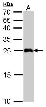
GSTA1 antibody detects GSTA1 protein by Western blot analysis. A. 50 μg mouse liver lysate/extract 12 % SDS-PAGE GSTA1 antibody (GTX108012) dilution: 1:5000
 antibody at 1:250 dilution.
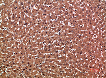
Antigen Retrieval: Trilogy? (EDTA based, pH 8.0) buffer, 15min")
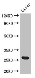
 A: Hep G2 (GTX27900) 12% SDS PAGE GSTA1 antibody GTX108012 diluted at 1:1000")
 antibody at 1:500 dilution.")
GSTA1 antibody detects GSTA1 protein by Western blot analysis. A. 50 μg mouse liver lysate/extract 12 % SDS-PAGE GSTA1 antibody (GTX108012) dilution: 1:5000
GSTA1 antibody
GTX108012
ApplicationsImmunoFluorescence, Western Blot, ImmunoCytoChemistry, ImmunoHistoChemistry, ImmunoHistoChemistry Paraffin
Product group Antibodies
ReactivityHuman, Mouse
TargetGSTA1
Overview
- SupplierGeneTex
- Product NameGSTA1 antibody
- Delivery Days Customer9
- Application Supplier NoteWB: 1:1000-1:10000. ICC/IF: 1:100-1:1000. IHC-P: 1:100-1:1000. *Optimal dilutions/concentrations should be determined by the researcher.Not tested in other applications.
- ApplicationsImmunoFluorescence, Western Blot, ImmunoCytoChemistry, ImmunoHistoChemistry, ImmunoHistoChemistry Paraffin
- CertificationResearch Use Only
- ClonalityPolyclonal
- Concentration6 mg/ml
- ConjugateUnconjugated
- Gene ID2938
- Target nameGSTA1
- Target descriptionglutathione S-transferase alpha 1
- Target synonymsGST-epsilon, GST2, GSTA1-1, GTH1, glutathione S-transferase A1, 13-hydroperoxyoctadecadienoate peroxidase, GST HA subunit 1, GST class-alpha member 1, S-(hydroxyalkyl)glutathione lyase A1, androst-5-ene-3,17-dione isomerase, glutathione S-alkyltransferase A1, glutathione S-aryltransferase A1, glutathione S-transferase 2, glutathione S-transferase Ha subunit 1, testicular tissue protein Li 80
- HostRabbit
- IsotypeIgG
- Protein IDP08263
- Protein NameGlutathione S-transferase A1
- Scientific DescriptionCytosolic and membrane-bound forms of glutathione S-transferase are encoded by two distinct supergene families. These enzymes function in the detoxification of electrophilic compounds, including carcinogens, therapeutic drugs, environmental toxins and products of oxidative stress, by conjugation with glutathione. The genes encoding these enzymes are known to be highly polymorphic. These genetic variations can change an individuals susceptibility to carcinogens and toxins as well as affect the toxicity and efficacy of some drugs. At present, eight distinct classes of the soluble cytoplasmic mammalian glutathione S-transferases have been identified: alpha, kappa, mu, omega, pi, sigma, theta and zeta. This gene encodes a glutathione S-tranferase belonging to the alpha class. The alpha class genes, located in a cluster mapped to chromosome 6, are the most abundantly expressed glutathione S-transferases in liver. In addition to metabolizing bilirubin and certain anti-cancer drugs in the liver, the alpha class of these enzymes exhibit glutathione peroxidase activity thereby protecting the cells from reactive oxygen species and the products of peroxidation. [provided by RefSeq]
- ReactivityHuman, Mouse
- Storage Instruction-20°C or -80°C,2°C to 8°C
- UNSPSC41116161
Datasheet
Related products
Product group Antibodies
Anti-GSTA1 AntibodyA100673
ApplicationsELISA, ImmunoHistoChemistry
ReactivityHuman
- SizePrice
Product group Antibodies
Anti-GSTA1 Antibody144-01628
ApplicationsWestern Blot
ReactivityHuman, Mouse
TargetGSTA1
- SizePrice
Product group Antibodies

GSTA1 AntibodyLS-C831554
ApplicationsWestern Blot, ImmunoHistoChemistry
ReactivityHuman, Mouse, Rat
TargetGSTA1
- SizePrice

Product group Antibodies
Anti-GSTA1/A2/A3/A4/A5 Antibody Picoband(r)A01462-1-CARRIER-FREE
ApplicationsWestern Blot, ELISA, ImmunoHistoChemistry
ReactivityHuman, Mouse, Rat
TargetGSTA1
- SizePrice
Product group Antibodies
GSTA1 (6T5) Monoclonal AntibodyBSM-51083M
ApplicationsWestern Blot
ReactivityMouse
TargetGSTA1
- SizePrice
Product group Antibodies
GSTA1 AntibodyCSB-PA009970HA01HU
ApplicationsImmunoFluorescence, Western Blot, ELISA, ImmunoHistoChemistry
ReactivityHuman, Mouse
TargetGSTA1
- SizePrice
Product group Antibodies
GSTA1 Polyclonal AntibodyCAC14589
ApplicationsImmunoFluorescence, Western Blot, ELISA, ImmunoHistoChemistry
ReactivityMouse
TargetGSTA1
- SizePrice

Product group Antibodies
Anti-GSTA1 AntibodyHPA048934
ApplicationsImmunoHistoChemistry
ReactivityHuman
TargetGSTA1
- SizePrice
Product group Antibodies
GSTA1 antibodyGTX113722
ApplicationsImmunoFluorescence, Western Blot, ImmunoCytoChemistry, ImmunoHistoChemistry, ImmunoHistoChemistry Paraffin
ReactivityHuman, Mouse
TargetGSTA1
- SizePrice