


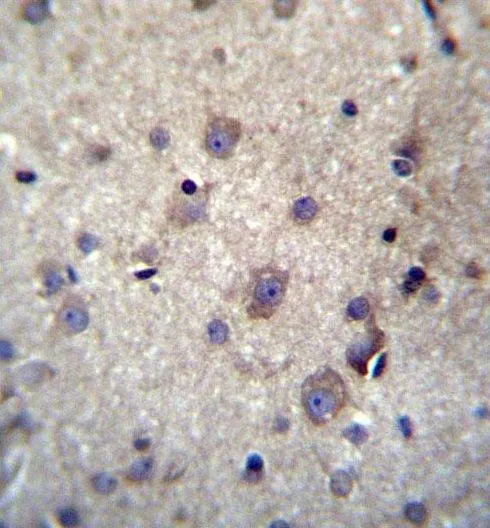
Immunohistochemistry analysis in human bone marrow and colon tissues using Anti-HBQ1 antibody. Corresponding HBQ1 RNA-seq data are presented for the same tissues.

Immunohistochemistry analysis in human bone marrow and colon tissues using Anti-HBQ1 antibody. Corresponding HBQ1 RNA-seq data are presented for the same tissues.
Anti-HBQ1 Antibody
HPA062473
ApplicationsImmunoCytoChemistry, ImmunoHistoChemistry
Product group Antibodies
ReactivityHuman
TargetHBQ1
Overview
- SupplierAtlas Antibodies
- Product NameAnti-HBQ1 Antibody
- Delivery Days Customer4
- ApplicationsImmunoCytoChemistry, ImmunoHistoChemistry
- CertificationResearch Use Only
- ClonalityPolyclonal
- ConjugateUnconjugated
- Gene ID3049
- Target nameHBQ1
- Target descriptionhemoglobin subunit theta 1
- Target synonymsHBQ, hemoglobin subunit theta-1, hemoglobin theta-1 chain, hemoglobin, theta 1, theta-1-globin
- HostRabbit
- IsotypeIgG
- Protein IDP09105
- Protein NameHemoglobin subunit theta-1
- Scientific DescriptionRecombinant Protein Epitope Signature Tag (PrEST) antigen sequence
- ReactivityHuman
- Storage Instruction-20°C,2°C to 8°C
- UNSPSC41116161
Datasheet
MSDS
Related products

Product group Antibodies
Anti-HBQ1 Antibody Picoband(r)A14380-1-CARRIER-FREE
ApplicationsWestern Blot, ELISA
ReactivityHuman, Mouse, Rat
TargetHBQ1
- SizePrice
Product group Antibodies
HBQ1 Polyclonal AntibodyBS-15419R
ApplicationsImmunoFluorescence, Western Blot, ELISA, ImmunoCytoChemistry, ImmunoHistoChemistry, ImmunoHistoChemistry Frozen, ImmunoHistoChemistry Paraffin
ReactivityHuman
TargetHBQ1
- SizePrice
Product group Antibodies
HBQ1 AntibodyCSB-PA010159LA01HU
ApplicationsELISA
ReactivityHuman
TargetHBQ1
- SizePrice
Product group Antibodies

HBQ1 Antibody (HRP)LS-C375879
ApplicationsELISA
ReactivityHuman
TargetHBQ1
- SizePrice
Product group Antibodies
Hemoglobin theta 1 antibodyGTX53555
ApplicationsWestern Blot, ImmunoHistoChemistry, ImmunoHistoChemistry Paraffin
ReactivityHuman
TargetHBQ1
- SizePrice