

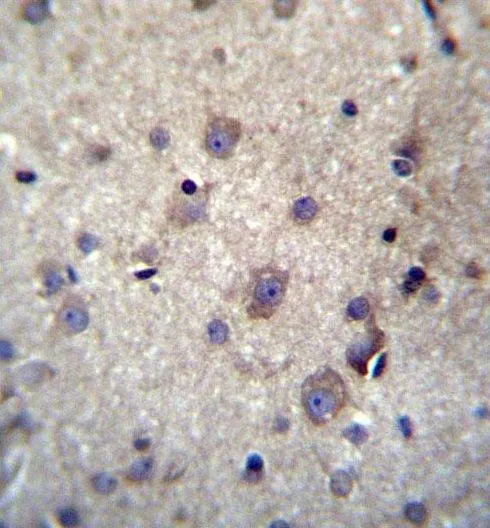
IHC-P analysis of human brain tissue using GTX53555 Hemoglobin theta 1 antibody.
 using GTX53555 Hemoglobin theta 1 antibody.")
IHC-P analysis of human brain tissue using GTX53555 Hemoglobin theta 1 antibody.
Hemoglobin theta 1 antibody
GTX53555
ApplicationsWestern Blot, ImmunoHistoChemistry, ImmunoHistoChemistry Paraffin
Product group Antibodies
ReactivityHuman
TargetHBQ1
Overview
- SupplierGeneTex
- Product NameHemoglobin theta 1 antibody
- Delivery Days Customer9
- Application Supplier NoteWB: 1:1000. IHC-P: 1:10-1:50. *Optimal dilutions/concentrations should be determined by the researcher.Not tested in other applications.
- ApplicationsWestern Blot, ImmunoHistoChemistry, ImmunoHistoChemistry Paraffin
- CertificationResearch Use Only
- ClonalityPolyclonal
- ConjugateUnconjugated
- Gene ID3049
- Target nameHBQ1
- Target descriptionhemoglobin subunit theta 1
- Target synonymsHBQ, hemoglobin subunit theta-1, hemoglobin theta-1 chain, hemoglobin, theta 1, theta-1-globin
- HostRabbit
- IsotypeIgG
- Protein IDP09105
- Protein NameHemoglobin subunit theta-1
- Scientific DescriptionTheta-globin mRNA is found in human fetal erythroid tissue but not in adult erythroid or other nonerythroid tissue. The theta-1 gene may be expressed very early in embryonic life, perhaps sometime before 5 weeks. Theta-1 is a member of the human alpha-globin gene cluster that involves five functional genes and two pseudogenes. The order of genes is: 5 - zeta - pseudozeta - mu - pseudoalpha-2 -pseudoalpha-1 - alpha-2 - alpha-1 - theta-1 - 3. Research supports a transcriptionally active role for the gene and a functional role for the peptide in specific cells, possibly those of early erythroid tissue. [provided by RefSeq, Jul 2008]
- ReactivityHuman
- Storage Instruction-20°C or -80°C,2°C to 8°C
- UNSPSC41116161
Datasheet
Related products

Product group Antibodies
Anti-HBQ1 Antibody Picoband(r)A14380-1-CARRIER-FREE
ApplicationsWestern Blot, ELISA
ReactivityHuman, Mouse, Rat
TargetHBQ1
- SizePrice
Product group Antibodies
HBQ1 AntibodyCSB-PA010159LA01HU
ApplicationsELISA
ReactivityHuman
TargetHBQ1
- SizePrice

Product group Antibodies
Anti-HBQ1 AntibodyHPA062473
ApplicationsImmunoCytoChemistry, ImmunoHistoChemistry
ReactivityHuman
TargetHBQ1
- SizePrice
Product group Antibodies

HBQ1 Antibody (HRP)LS-C375879
ApplicationsELISA
ReactivityHuman
TargetHBQ1
- SizePrice
Product group Antibodies
HBQ1 Polyclonal AntibodyBS-15419R
ApplicationsImmunoFluorescence, Western Blot, ELISA, ImmunoCytoChemistry, ImmunoHistoChemistry, ImmunoHistoChemistry Frozen, ImmunoHistoChemistry Paraffin
ReactivityHuman
TargetHBQ1
- SizePrice