


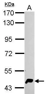
Figure 1. Western blot analysis of HERP/HERPUD1 using anti-HERP/HERPUD1 antibody (A04908-2). Electrophoresis was performed on a 5-20% SDS-PAGE gel at 70V (Stacking gel) / 90V (Resolving gel) for 2-3 hours. The sample well of each lane was loaded with 30 ug of sample under reducing conditions. Lane 1: human PC-3 whole cell lysates, Lane 2: human Hela whole cell lysates, Lane 3: human HepG2 whole cell lysates, Lane 4: human LNCAP whole cell lysates, Lane 5: rat liver tissue lysates, Lane 6: mouse liver tissue lysates. After electrophoresis, proteins were transferred to a nitrocellulose membrane at 150 mA for 50-90 minutes. Blocked the membrane with 5% non-fat milk/TBS for 1.5 hour at RT. The membrane was incubated with rabbit anti-HERP/HERPUD1 antigen affinity purified polyclonal antibody (Catalog # A04908-2) at 0.5 microg/mL overnight at 4°C, then washed with TBS-0.1%Tween 3 times with 5 minutes each and probed with a goat anti-rabbit IgG-HRP secondary antibody at a dilution of 1:5000 for 1.5 hour at RT. The signal is developed using an Enhanced Chemiluminescent detection (ECL) kit (Catalog # EK1002) with Tanon 5200 system. A specific band was detected for HERP/HERPUD1 at approximately 56 kDa. The expected band size for HERP/HERPUD1 is at 44 kDa.
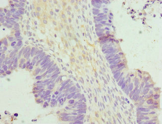
. HERP/HERPUD1 was detected in a paraffin-embedded section of human appendiceal carcinoid tissue. Heat mediated antigen retrieval was performed in EDTA buffer (pH 8.0, epitope retrieval solution). The tissue section was blocked with 10% goat serum. The tissue section was then incubated with 2 microg/ml rabbit anti-HERP/HERPUD1 Antibody (A04908-2) overnight at 4°C. Peroxidase Conjugated Goat Anti-rabbit IgG was used as secondary antibody and incubated for 30 minutes at 37°C. The tissue section was developed using HRP Conjugated Rabbit IgG Super Vision Assay Kit (Catalog # SV0002) with DAB as the chromogen.")
. HERP/HERPUD1 was detected in a paraffin-embedded section of human appendiceal carcinoid tissue. Heat mediated antigen retrieval was performed in EDTA buffer (pH 8.0, epitope retrieval solution). The tissue section was blocked with 10% goat serum. The tissue section was then incubated with 2 microg/ml rabbit anti-HERP/HERPUD1 Antibody (A04908-2) overnight at 4°C. Peroxidase Conjugated Goat Anti-rabbit IgG was used as secondary antibody and incubated for 30 minutes at 37°C. The tissue section was developed using HRP Conjugated Rabbit IgG Super Vision Assay Kit (Catalog # SV0002) with DAB as the chromogen.")
. HERP/HERPUD1 was detected in a paraffin-embedded section of human breast cancer tissue. Heat mediated antigen retrieval was performed in EDTA buffer (pH 8.0, epitope retrieval solution). The tissue section was blocked with 10% goat serum. The tissue section was then incubated with 2 microg/ml rabbit anti-HERP/HERPUD1 Antibody (A04908-2) overnight at 4°C. Peroxidase Conjugated Goat Anti-rabbit IgG was used as secondary antibody and incubated for 30 minutes at 37°C. The tissue section was developed using HRP Conjugated Rabbit IgG Super Vision Assay Kit (Catalog # SV0002) with DAB as the chromogen.")
. HERP/HERPUD1 was detected in a paraffin-embedded section of human breast cancer tissue. Heat mediated antigen retrieval was performed in EDTA buffer (pH 8.0, epitope retrieval solution). The tissue section was blocked with 10% goat serum. The tissue section was then incubated with 2 microg/ml rabbit anti-HERP/HERPUD1 Antibody (A04908-2) overnight at 4°C. Peroxidase Conjugated Goat Anti-rabbit IgG was used as secondary antibody and incubated for 30 minutes at 37°C. The tissue section was developed using HRP Conjugated Rabbit IgG Super Vision Assay Kit (Catalog # SV0002) with DAB as the chromogen.")
. HERP/HERPUD1 was detected in a paraffin-embedded section of human cervix squamous cell carcinoma tissue. Heat mediated antigen retrieval was performed in EDTA buffer (pH 8.0, epitope retrieval solution). The tissue section was blocked with 10% goat serum. The tissue section was then incubated with 2 microg/ml rabbit anti-HERP/HERPUD1 Antibody (A04908-2) overnight at 4°C. Peroxidase Conjugated Goat Anti-rabbit IgG was used as secondary antibody and incubated for 30 minutes at 37°C. The tissue section was developed using HRP Conjugated Rabbit IgG Super Vision Assay Kit (Catalog # SV0002) with DAB as the chromogen.")
. HERP/HERPUD1 was detected in a paraffin-embedded section of human cervix squamous cell carcinoma tissue. Heat mediated antigen retrieval was performed in EDTA buffer (pH 8.0, epitope retrieval solution). The tissue section was blocked with 10% goat serum. The tissue section was then incubated with 2 microg/ml rabbit anti-HERP/HERPUD1 Antibody (A04908-2) overnight at 4°C. Peroxidase Conjugated Goat Anti-rabbit IgG was used as secondary antibody and incubated for 30 minutes at 37°C. The tissue section was developed using HRP Conjugated Rabbit IgG Super Vision Assay Kit (Catalog # SV0002) with DAB as the chromogen.")
. HERP/HERPUD1 was detected in a paraffin-embedded section of human ovarian cancer tissue. Heat mediated antigen retrieval was performed in EDTA buffer (pH 8.0, epitope retrieval solution). The tissue section was blocked with 10% goat serum. The tissue section was then incubated with 2 microg/ml rabbit anti-HERP/HERPUD1 Antibody (A04908-2) overnight at 4°C. Peroxidase Conjugated Goat Anti-rabbit IgG was used as secondary antibody and incubated for 30 minutes at 37°C. The tissue section was developed using HRP Conjugated Rabbit IgG Super Vision Assay Kit (Catalog # SV0002) with DAB as the chromogen.")
. HERP/HERPUD1 was detected in a paraffin-embedded section of human ovarian cancer tissue. Heat mediated antigen retrieval was performed in EDTA buffer (pH 8.0, epitope retrieval solution). The tissue section was blocked with 10% goat serum. The tissue section was then incubated with 2 microg/ml rabbit anti-HERP/HERPUD1 Antibody (A04908-2) overnight at 4°C. Peroxidase Conjugated Goat Anti-rabbit IgG was used as secondary antibody and incubated for 30 minutes at 37°C. The tissue section was developed using HRP Conjugated Rabbit IgG Super Vision Assay Kit (Catalog # SV0002) with DAB as the chromogen.")
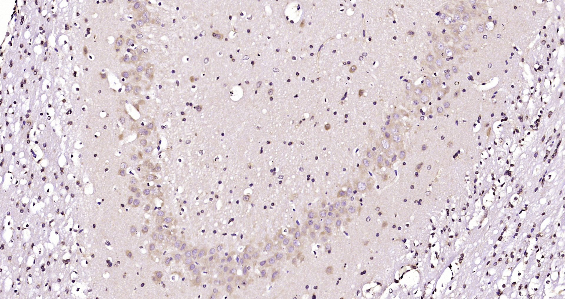
. HERP/HERPUD1 was detected in a paraffin-embedded section of human rectum adenocarcinoma tissue. Heat mediated antigen retrieval was performed in EDTA buffer (pH 8.0, epitope retrieval solution). The tissue section was blocked with 10% goat serum. The tissue section was then incubated with 2 microg/ml rabbit anti-HERP/HERPUD1 Antibody (A04908-2) overnight at 4°C. Peroxidase Conjugated Goat Anti-rabbit IgG was used as secondary antibody and incubated for 30 minutes at 37°C. The tissue section was developed using HRP Conjugated Rabbit IgG Super Vision Assay Kit (Catalog # SV0002) with DAB as the chromogen.")
Figure 1. Western blot analysis of HERP/HERPUD1 using anti-HERP/HERPUD1 antibody (A04908-2). Electrophoresis was performed on a 5-20% SDS-PAGE gel at 70V (Stacking gel) / 90V (Resolving gel) for 2-3 hours. The sample well of each lane was loaded with 30 ug of sample under reducing conditions. Lane 1: human PC-3 whole cell lysates, Lane 2: human Hela whole cell lysates, Lane 3: human HepG2 whole cell lysates, Lane 4: human LNCAP whole cell lysates, Lane 5: rat liver tissue lysates, Lane 6: mouse liver tissue lysates. After electrophoresis, proteins were transferred to a nitrocellulose membrane at 150 mA for 50-90 minutes. Blocked the membrane with 5% non-fat milk/TBS for 1.5 hour at RT. The membrane was incubated with rabbit anti-HERP/HERPUD1 antigen affinity purified polyclonal antibody (Catalog # A04908-2) at 0.5 microg/mL overnight at 4°C, then washed with TBS-0.1%Tween 3 times with 5 minutes each and probed with a goat anti-rabbit IgG-HRP secondary antibody at a dilution of 1:5000 for 1.5 hour at RT. The signal is developed using an Enhanced Chemiluminescent detection (ECL) kit (Catalog # EK1002) with Tanon 5200 system. A specific band was detected for HERP/HERPUD1 at approximately 56 kDa. The expected band size for HERP/HERPUD1 is at 44 kDa.
Anti-HERP/HERPUD1 Antibody Picoband(r)
A04908-2-CARRIER-FREE
ApplicationsImmunoFluorescence, Western Blot, ELISA, ImmunoCytoChemistry, ImmunoHistoChemistry
Product group Antibodies
ReactivityHuman, Mouse, Rat
TargetHERPUD1
Overview
- SupplierBoster Bio
- Product NameAnti-HERP/HERPUD1 Antibody Picoband(r)
- Delivery Days Customer9
- ApplicationsImmunoFluorescence, Western Blot, ELISA, ImmunoCytoChemistry, ImmunoHistoChemistry
- CertificationResearch Use Only
- ClonalityPolyclonal
- Concentration500 ug/ml
- Gene ID9709
- Target nameHERPUD1
- Target descriptionhomocysteine inducible ER protein with ubiquitin like domain 1
- Target synonymsHERP, HERPUD1-IT1, Mif1, SUP, homocysteine-responsive endoplasmic reticulum-resident ubiquitin-like domain member 1 protein, HERPUD1 intronic transcript 1, MMS-inducible, homocysteine-inducible endoplasmic reticulum stress-inducible ubiquitin-like domain member 1 protein, homocysteine-inducible, endoplasmic reticulum stress-inducible, ubiquitin-like domain member 1, methyl methanesulfonate (MMF)-inducible fragment protein 1
- HostRabbit
- IsotypeIgG
- Protein IDQ15011
- Protein NameHomocysteine-responsive endoplasmic reticulum-resident ubiquitin-like domain member 1 protein
- Scientific DescriptionBoster Bio Anti-HERP/HERPUD1 Antibody Picoband® catalog # A07951-1. Tested in WB, IHC, ICC/IF, IF, ELISA applications. This antibody reacts with Human, Mouse, Rat. The brand Picoband indicates this is a premium antibody that guarantees superior quality, high affinity, and strong signals with minimal background in Western blot applications. Only our best-performing antibodies are designated as Picoband, ensuring unmatched performance.
- ReactivityHuman, Mouse, Rat
- Storage Instruction-20°C,2°C to 8°C
- UNSPSC12352203
Related products
Product group Antibodies
Anti-HERPUD1 (N-term) Antibody102-22649
ApplicationsWestern Blot
TargetHERPUD1
- SizePrice
Product group Antibodies
HERPUD1 Polyclonal AntibodyBS-15464R
ApplicationsImmunoFluorescence, Western Blot, ELISA, ImmunoCytoChemistry, ImmunoHistoChemistry, ImmunoHistoChemistry Frozen, ImmunoHistoChemistry Paraffin
ReactivityBovine, Equine, Human, Mouse, Porcine, Rabbit, Rat, Sheep
TargetHERPUD1
- SizePrice
Product group Antibodies

HERPUD1 / HERP AntibodyLS-C749110
ApplicationsWestern Blot, ImmunoHistoChemistry
ReactivityHuman
TargetHERPUD1
- SizePrice
Product group Antibodies
Anti-HERPUD1 AntibodyA31837
ApplicationsWestern Blot, ImmunoHistoChemistry
ReactivityHuman
- SizePrice

Product group Antibodies
Anti-HERPUD1 AntibodyHPA040754
ApplicationsWestern Blot, ImmunoHistoChemistry
ReactivityHuman
TargetHERPUD1
- SizePrice
Product group Antibodies
HERPUD1 AntibodyCSB-PA622984ESR1HU
ApplicationsELISA, ImmunoHistoChemistry
ReactivityHuman
TargetHERPUD1
- SizePrice
Product group Antibodies
HERPUD1 antibodyGTX112261
ApplicationsWestern Blot, ImmunoHistoChemistry, ImmunoHistoChemistry Paraffin
ReactivityHuman, Mouse
TargetHERPUD1
- SizePrice