


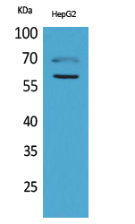
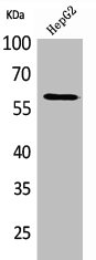
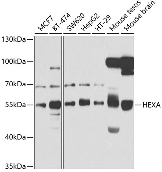
Figure 1. Western blot analysis of HEXA using anti-HEXA antibody (A00692-1). Electrophoresis was performed on a 5-20% SDS-PAGE gel at 70V (Stacking gel) / 90V (Resolving gel) for 2-3 hours. The sample well of each lane was loaded with 30 ug of sample under reducing conditions. Lane 1: human 293T whole cell lysates, Lane 2: human Caco-2 whole cell lysates, Lane 3: human Hela whole cell lysates, Lane 4: rat kidney tissue lysates, Lane 5: mouse kidney tissue lysates. After electrophoresis, proteins were transferred to a nitrocellulose membrane at 150 mA for 50-90 minutes. Blocked the membrane with 5% non-fat milk/TBS for 1.5 hour at RT. The membrane was incubated with rabbit anti-HEXA antigen affinity purified polyclonal antibody (Catalog # A00692-1) at 0.25 microg/mL overnight at 4°C, then washed with TBS-0.1%Tween 3 times with 5 minutes each and probed with a goat anti-rabbit IgG-HRP secondary antibody at a dilution of 1:5000 for 1.5 hour at RT. The signal is developed using an Enhanced Chemiluminescent detection (ECL) kit (Catalog # EK1002) with Tanon 5200 system. A specific band was detected for HEXA at approximately 60 kDa. The expected band size for HEXA is at 60 kDa.
. HEXA was detected in an immunocytochemical section of U20S cells. Enzyme antigen retrieval was performed using IHC enzyme antigen retrieval reagent (AR0022) for 15 mins. The cells were blocked with 10% goat serum. And then incubated with 5 microg/mL rabbit anti-HEXA Antibody (A00692-1) overnight at 4°C. DyLight®488 Conjugated Goat Anti-Rabbit IgG (BA1127) was used as secondary antibody at 1:100 dilution and incubated for 30 minutes at 37°C. The section was counterstained with DAPI. Visualize using a fluorescence microscope and filter sets appropriate for the label used.")
Figure 1. Western blot analysis of HEXA using anti-HEXA antibody (A00692-1). Electrophoresis was performed on a 5-20% SDS-PAGE gel at 70V (Stacking gel) / 90V (Resolving gel) for 2-3 hours. The sample well of each lane was loaded with 30 ug of sample under reducing conditions. Lane 1: human 293T whole cell lysates, Lane 2: human Caco-2 whole cell lysates, Lane 3: human Hela whole cell lysates, Lane 4: rat kidney tissue lysates, Lane 5: mouse kidney tissue lysates. After electrophoresis, proteins were transferred to a nitrocellulose membrane at 150 mA for 50-90 minutes. Blocked the membrane with 5% non-fat milk/TBS for 1.5 hour at RT. The membrane was incubated with rabbit anti-HEXA antigen affinity purified polyclonal antibody (Catalog # A00692-1) at 0.25 microg/mL overnight at 4°C, then washed with TBS-0.1%Tween 3 times with 5 minutes each and probed with a goat anti-rabbit IgG-HRP secondary antibody at a dilution of 1:5000 for 1.5 hour at RT. The signal is developed using an Enhanced Chemiluminescent detection (ECL) kit (Catalog # EK1002) with Tanon 5200 system. A specific band was detected for HEXA at approximately 60 kDa. The expected band size for HEXA is at 60 kDa.
Anti-HEXA Antibody Picoband(r)
A00692-1-CARRIER-FREE
ApplicationsImmunoFluorescence, Western Blot, ELISA, ImmunoCytoChemistry
Product group Antibodies
ReactivityHuman, Mouse, Rat
TargetHEXA
Overview
- SupplierBoster Bio
- Product NameAnti-HEXA Antibody Picoband(r)
- Delivery Days Customer9
- Application Supplier NoteTested Species: In-house tested species with positive results. Other applications have not been tested. Optimal dilutions should be determined by end users.
- ApplicationsImmunoFluorescence, Western Blot, ELISA, ImmunoCytoChemistry
- CertificationResearch Use Only
- ClonalityPolyclonal
- Concentration500 ug/ml
- Gene ID3073
- Target nameHEXA
- Target descriptionhexosaminidase subunit alpha
- Target synonymsTSD, beta-hexosaminidase subunit alpha, N-acetyl-beta-glucosaminidase subunit alpha, beta-N-acetylhexosaminidase subunit alpha, hexosaminidase A (alpha polypeptide), hexosaminidase subunit A
- HostRabbit
- IsotypeIgG
- Protein IDP06865
- Protein NameBeta-hexosaminidase subunit alpha
- Scientific DescriptionBoster Bio Anti-HEXA Antibody Picoband® catalog # A00692-1. Tested in ELISA, IF, ICC, WB applications. This antibody reacts with Human, Mouse, Rat. The brand Picoband indicates this is a premium antibody that guarantees superior quality, high affinity, and strong signals with minimal background in Western blot applications. Only our best-performing antibodies are designated as Picoband, ensuring unmatched performance.
- ReactivityHuman, Mouse, Rat
- Storage Instruction-20°C,2°C to 8°C
- UNSPSC12352203
Related products
Product group Antibodies
Anti-HEXA Antibody144-65959
ApplicationsWestern Blot
ReactivityHuman, Mouse
TargetHEXA
- SizePrice
Product group Antibodies
Anti-HEXA AntibodyA40722
ApplicationsWestern Blot, ELISA
ReactivityHuman, Mouse, Rat
- SizePrice
Product group Antibodies
Hexa Polyclonal AntibodyCAC11280
ApplicationsImmunoFluorescence, Western Blot, ELISA, ImmunoHistoChemistry
ReactivityRat
TargetHEXA
- SizePrice
Product group Antibodies
HEXA AntibodyCSB-PA005178
ApplicationsWestern Blot, ELISA
ReactivityHuman, Mouse, Rat
TargetHEXA
- SizePrice
Product group Antibodies
HEXA antibodyGTX33239
ApplicationsWestern Blot
ReactivityHuman, Mouse
TargetHEXA
- SizePrice

Product group Antibodies
Anti-HEXA AntibodyHPA018082
ApplicationsImmunoCytoChemistry
ReactivityHuman
TargetHEXA
- SizePrice
Product group Antibodies

HEXA AntibodyLS-C747545
ApplicationsWestern Blot, ImmunoHistoChemistry
ReactivityHuman, Mouse, Rat
TargetHEXA
- SizePrice
Product group Antibodies
Anti-HEXA AntibodyCAB5646
ApplicationsWestern Blot, ELISA
ReactivityHuman
TargetHEXA
- SizePrice