


Immunofluorescent staining of human cell line SK-MEL-30 shows localization to vesicles.
Immunofluorescent staining of human cell line SK-MEL-30 shows localization to vesicles.
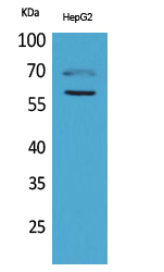
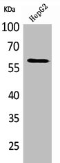
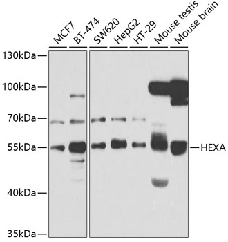
Anti-HEXA Antibody
HPA018082
ApplicationsImmunoCytoChemistry
Product group Antibodies
ReactivityHuman
TargetHEXA
Overview
- SupplierAtlas Antibodies
- Product NameAnti-HEXA Antibody
- Delivery Days Customer4
- ApplicationsImmunoCytoChemistry
- CertificationResearch Use Only
- ClonalityPolyclonal
- ConjugateUnconjugated
- Gene ID3073
- Target nameHEXA
- Target descriptionhexosaminidase subunit alpha
- Target synonymsTSD, beta-hexosaminidase subunit alpha, N-acetyl-beta-glucosaminidase subunit alpha, beta-N-acetylhexosaminidase subunit alpha, hexosaminidase A (alpha polypeptide), hexosaminidase subunit A
- HostRabbit
- IsotypeIgG
- Protein IDP06865
- Protein NameBeta-hexosaminidase subunit alpha
- Scientific DescriptionRecombinant Protein Epitope Signature Tag (PrEST) antigen sequence
- ReactivityHuman
- Storage Instruction-20°C,2°C to 8°C
- UNSPSC41116161
Datasheet
MSDS
Related products

Product group Antibodies
Anti-HEXA Antibody Picoband(r)A00692-1-CARRIER-FREE
ApplicationsImmunoFluorescence, Western Blot, ELISA, ImmunoCytoChemistry
ReactivityHuman, Mouse, Rat
TargetHEXA
- SizePrice
Product group Antibodies
Anti-HEXA Antibody144-65959
ApplicationsWestern Blot
ReactivityHuman, Mouse
TargetHEXA
- SizePrice
Product group Antibodies
Anti-HEXA AntibodyA40722
ApplicationsWestern Blot, ELISA
ReactivityHuman, Mouse, Rat
- SizePrice
Product group Antibodies
Hexa Polyclonal AntibodyCAC11280
ApplicationsImmunoFluorescence, Western Blot, ELISA, ImmunoHistoChemistry
ReactivityRat
TargetHEXA
- SizePrice
Product group Antibodies
HEXA AntibodyCSB-PA005178
ApplicationsWestern Blot, ELISA
ReactivityHuman, Mouse, Rat
TargetHEXA
- SizePrice
Product group Antibodies
HEXA antibodyGTX33239
ApplicationsWestern Blot
ReactivityHuman, Mouse
TargetHEXA
- SizePrice
Product group Antibodies

HEXA AntibodyLS-C747545
ApplicationsWestern Blot, ImmunoHistoChemistry
ReactivityHuman, Mouse, Rat
TargetHEXA
- SizePrice
Product group Antibodies
Anti-HEXA AntibodyCAB5646
ApplicationsWestern Blot, ELISA
ReactivityHuman
TargetHEXA
- SizePrice