


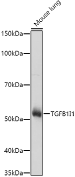
Figure 1. Western blot analysis of HIC5/TGFB1I1 using anti-HIC5/TGFB1I1 antibody (A04630-2). Electrophoresis was performed on a 5-20% SDS-PAGE gel at 70V (Stacking gel) / 90V (Resolving gel) for 2-3 hours. The sample well of each lane was loaded with 30 ug of sample under reducing conditions. Lane 1: monkey COS-7 whole cell lysates. After electrophoresis, proteins were transferred to a nitrocellulose membrane at 150 mA for 50-90 minutes. Blocked the membrane with 5% non-fat milk/TBS for 1.5 hour at RT. The membrane was incubated with rabbit anti-HIC5/TGFB1I1 antigen affinity purified polyclonal antibody (Catalog # A04630-2) at 0.5 microg/mL overnight at 4°C, then washed with TBS-0.1%Tween 3 times with 5 minutes each and probed with a goat anti-rabbit IgG-HRP secondary antibody at a dilution of 1:5000 for 1.5 hour at RT. The signal is developed using an Enhanced Chemiluminescent detection (ECL) kit (Catalog # EK1002) with Tanon 5200 system. A specific band was detected for HIC5/TGFB1I1 at approximately 75 kDa. The expected band size for HIC5/TGFB1I1 is at 75 kDa.
Figure 1. Western blot analysis of HIC5/TGFB1I1 using anti-HIC5/TGFB1I1 antibody (A04630-2). Electrophoresis was performed on a 5-20% SDS-PAGE gel at 70V (Stacking gel) / 90V (Resolving gel) for 2-3 hours. The sample well of each lane was loaded with 30 ug of sample under reducing conditions. Lane 1: monkey COS-7 whole cell lysates. After electrophoresis, proteins were transferred to a nitrocellulose membrane at 150 mA for 50-90 minutes. Blocked the membrane with 5% non-fat milk/TBS for 1.5 hour at RT. The membrane was incubated with rabbit anti-HIC5/TGFB1I1 antigen affinity purified polyclonal antibody (Catalog # A04630-2) at 0.5 microg/mL overnight at 4°C, then washed with TBS-0.1%Tween 3 times with 5 minutes each and probed with a goat anti-rabbit IgG-HRP secondary antibody at a dilution of 1:5000 for 1.5 hour at RT. The signal is developed using an Enhanced Chemiluminescent detection (ECL) kit (Catalog # EK1002) with Tanon 5200 system. A specific band was detected for HIC5/TGFB1I1 at approximately 75 kDa. The expected band size for HIC5/TGFB1I1 is at 75 kDa.
Anti-HIC5/TGFB1I1 Antibody Picoband(r)
A04630-2-CARRIER-FREE
ApplicationsWestern Blot, ELISA
Product group Antibodies
TargetTGFB1I1
Overview
- SupplierBoster Bio
- Product NameAnti-HIC5/TGFB1I1 Antibody Picoband(r)
- Delivery Days Customer9
- ApplicationsWestern Blot, ELISA
- CertificationResearch Use Only
- ClonalityPolyclonal
- Concentration500 ug/ml
- Gene ID7041
- Target nameTGFB1I1
- Target descriptiontransforming growth factor beta 1 induced transcript 1
- Target synonymsARA55, HIC-5, HIC5, TSC-5, transforming growth factor beta-1-induced transcript 1 protein, androgen receptor coactivator 55 kDa protein, androgen receptor coactivator ARA55, androgen receptor-associated protein of 55 kDa, hydrogen peroxide-inducible clone 5 protein, hydrogen peroxide-inducible clone-5
- HostRabbit
- IsotypeIgG
- Protein IDO43294
- Protein NameTransforming growth factor beta-1-induced transcript 1 protein
- Scientific DescriptionBoster Bio Anti-HIC5/TGFB1I1 Antibody Picoband® catalog # A04630-2. Tested in ELISA, WB applications. This antibody reacts with Human, Monkey. The brand Picoband indicates this is a premium antibody that guarantees superior quality, high affinity, and strong signals with minimal background in Western blot applications. Only our best-performing antibodies are designated as Picoband, ensuring unmatched performance.
- Storage Instruction-20°C,2°C to 8°C
- UNSPSC12352203
Related products
Product group Antibodies
TGFB1I1 Polyclonal AntibodyCAC15009
ApplicationsWestern Blot, ELISA, ImmunoHistoChemistry
TargetTGFB1I1
- SizePrice
Product group Antibodies
Anti-Mouse TGFB1I1 Antibody144-08459
ApplicationsWestern Blot
TargetTGFB1I1
- SizePrice
Product group Antibodies
References
HIC5 antibody [N1], N-termGTX108510
ApplicationsImmunoFluorescence, Western Blot, ImmunoCytoChemistry
TargetTGFB1I1
- SizePrice
Product group Antibodies

ARA55 / HIC-5 AntibodyLS-C409994
ApplicationsWestern Blot
TargetTGFB1I1
- SizePrice
Product group Antibodies
TGFB1I1 AntibodyCSB-PA050247
ApplicationsImmunoFluorescence, Western Blot, ELISA, ImmunoHistoChemistry
ReactivityHuman, Mouse, Rat
TargetTGFB1I1
- SizePrice