


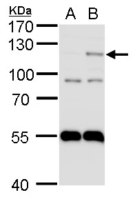
Figure 1. Western blot analysis of HIF-2-Alpha/EPAS1 using anti-HIF-2-Alpha/EPAS1 antibody (A01248-1). Electrophoresis was performed on a 5-20% SDS-PAGE gel at 70V (Stacking gel) / 90V (Resolving gel) for 2-3 hours. The sample well of each lane was loaded with 30ug of sample under reducing conditions. Lane 1: rat lung tissue lysates, Lane 2: rat PC-12 whole cell lysates, Lane 3: mouse lung tissue lysates, Lane 4: mouse SP2/0 whole cell lysates. After Electrophoresis, proteins were transferred to a Nitrocellulose membrane at 150mA for 50-90 minutes. Blocked the membrane with 5% Non-fat Milk/ TBS for 1.5 hour at RT. The membrane was incubated with rabbit anti-HIF-2-Alpha/EPAS1 antigen affinity purified polyclonal antibody (Catalog # A01248-1) at 0.5 microg/mL overnight at 4°C, then washed with TBS-0.1%Tween 3 times with 5 minutes each and probed with a goat anti-rabbit IgG-HRP secondary antibody at a dilution of 1:5000 for 1.5 hour at RT. The signal is developed using an Enhanced Chemiluminescent detection (ECL) kit (Catalog # EK1002) with Tanon 5200 system. A specific band was detected for HIF-2-Alpha/EPAS1 at approximately 96KD. The expected band size for HIF-2-Alpha/EPAS1 is at 96KD.
. Overlay histogram showing SiHa cells stained with A01248-1 (Blue line). To facilitate intracellular staining, cells were fixed with 4% paraformaldehyde and permeabilized with permeabilization buffer. The cells were blocked with 10% normal goat serum. And then incubated with rabbit anti-HIF-2-Alpha/EPAS1 Antibody (A01248-1, 1microg/1x106 cells) for 30 min at 20°C. DyLight®488 conjugated goat anti-rabbit IgG (BA1127, 5-10microg/1x106 cells) was used as secondary antibody for 30 minutes at 20°C. Isotype control antibody (Green line) was rabbit IgG (1microg/1x106) used under the same conditions. Unlabelled sample without incubation with primary antibody and secondary antibody (Red line) was used as a blank control.")
Figure 1. Western blot analysis of HIF-2-Alpha/EPAS1 using anti-HIF-2-Alpha/EPAS1 antibody (A01248-1). Electrophoresis was performed on a 5-20% SDS-PAGE gel at 70V (Stacking gel) / 90V (Resolving gel) for 2-3 hours. The sample well of each lane was loaded with 30ug of sample under reducing conditions. Lane 1: rat lung tissue lysates, Lane 2: rat PC-12 whole cell lysates, Lane 3: mouse lung tissue lysates, Lane 4: mouse SP2/0 whole cell lysates. After Electrophoresis, proteins were transferred to a Nitrocellulose membrane at 150mA for 50-90 minutes. Blocked the membrane with 5% Non-fat Milk/ TBS for 1.5 hour at RT. The membrane was incubated with rabbit anti-HIF-2-Alpha/EPAS1 antigen affinity purified polyclonal antibody (Catalog # A01248-1) at 0.5 microg/mL overnight at 4°C, then washed with TBS-0.1%Tween 3 times with 5 minutes each and probed with a goat anti-rabbit IgG-HRP secondary antibody at a dilution of 1:5000 for 1.5 hour at RT. The signal is developed using an Enhanced Chemiluminescent detection (ECL) kit (Catalog # EK1002) with Tanon 5200 system. A specific band was detected for HIF-2-Alpha/EPAS1 at approximately 96KD. The expected band size for HIF-2-Alpha/EPAS1 is at 96KD.
Anti-HIF-2-alpha/EPAS1 Antibody Picoband(r)
A01248-1-CARRIER-FREE
ApplicationsFlow Cytometry, Western Blot
Product group Antibodies
ReactivityHuman, Mouse, Rat
TargetEPAS1
Overview
- SupplierBoster Bio
- Product NameAnti-HIF-2-alpha/EPAS1 Antibody Picoband(r)
- Delivery Days Customer9
- ApplicationsFlow Cytometry, Western Blot
- CertificationResearch Use Only
- ClonalityPolyclonal
- Concentration500 ug/ml
- Gene ID2034
- Target nameEPAS1
- Target descriptionendothelial PAS domain protein 1
- Target synonymsECYT4, HIF2A, HLF, MOP2, PASD2, bHLHe73, endothelial PAS domain-containing protein 1, EPAS-1, HIF-1-alpha-like factor, HIF-1alpha-like factor, HIF-2-alpha, HIF2-alpha, PAS domain-containing protein 2, basic-helix-loop-helix-PAS protein MOP2, class E basic helix-loop-helix protein 73, hypoxia-inducible factor 2 alpha, member of PAS protein 2
- HostRabbit
- IsotypeIgG
- Protein IDQ99814
- Protein NameEndothelial PAS domain-containing protein 1
- Scientific DescriptionBoster Bio Anti-HIF-2-alpha/EPAS1 Antibody Picoband® catalog # A01248-1. Tested in Flow Cytometry, WB applications. This antibody reacts with Human, Mouse, Rat. The brand Picoband indicates this is a premium antibody that guarantees superior quality, high affinity, and strong signals with minimal background in Western blot applications. Only our best-performing antibodies are designated as Picoband, ensuring unmatched performance.
- ReactivityHuman, Mouse, Rat
- Storage Instruction-20°C,2°C to 8°C
- UNSPSC12352203
Related products
Product group Antibodies
EPAS1 AntibodyCSB-PA005478
ApplicationsWestern Blot, ELISA
ReactivityHuman, Mouse, Rat
TargetEPAS1
- SizePrice

Product group Antibodies
Anti-EPAS1 AntibodyHPA031200
ApplicationsImmunoCytoChemistry
ReactivityHuman
TargetEPAS1
- SizePrice
Product group Antibodies

HIF2A / EPAS1 AntibodyLS-C409163
ApplicationsImmunoFluorescence, Western Blot
ReactivityHuman
TargetEPAS1
- SizePrice
Product group Antibodies
ApplicationsImmunoPrecipitation, Western Blot, ImmunoCytoChemistry, ImmunoHistoChemistry
ReactivityMouse, Porcine
TargetEPAS1
- SizePrice
Product group Antibodies
HIF-2 alpha Recombinant AntibodyBSM-54278R
ApplicationsFlow Cytometry, ImmunoFluorescence, Western Blot, ImmunoCytoChemistry
ReactivityHuman, Mouse, Rat
TargetEPAS1
- SizePrice
Product group Antibodies
HIF2 alpha antibody [N1N3]GTX103707
ApplicationsWestern Blot, ChIP Chromatin ImmunoPrecipitation
ReactivityHuman, Zebra Fish
TargetEPAS1
- SizePrice
Product group Antibodies
Anti-HIF2a Antibody144-66426
ApplicationsImmunoFluorescence, Western Blot
ReactivityHuman, Mouse, Rat
TargetEPAS1
- SizePrice