


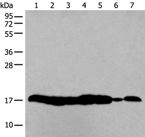
Figure 1. Western blot analysis of Histone H3 (mono methyl K18) using anti-Histone H3 (mono methyl K18) antibody (M12477-6). Electrophoresis was performed on a 5-20% SDS-PAGE gel at 70V (Stacking gel) / 90V (Resolving gel) for 2-3 hours. The sample well of each lane was loaded with 30 ug of sample under reducing conditions. Lane 1: human Hela whole cell lysates, Lane 2: human 293T whole cell lysates, Lane 3: human CACO-2 whole cell lysates, Lane 4: human HepG2 whole cell lysates, Lane 5: human PC-12 whole cell lysates, Lane 6: rat C6 whole cell lysates, Lane 7: mouse RAW264.7 whole cell lysates, Lane 8: mouse NIH/3T3 whole cell lysates. After electrophoresis, proteins were transferred to a nitrocellulose membrane at 150 mA for 50-90 minutes. Blocked the membrane with 5% non-fat milk/TBS for 1.5 hour at RT. The membrane was incubated with rabbit anti-Histone H3 (mono methyl K18) antigen affinity purified monoclonal antibody (Catalog # M12477-6) at 1:500 overnight at 4°C, then washed with TBS-0.1%Tween 3 times with 5 minutes each and probed with a goat anti-rabbit IgG-HRP secondary antibody at a dilution of 1:5000 for 1.5 hour at RT. The signal is developed using an Enhanced Chemiluminescent detection (ECL) kit (Catalog # EK1002) with Tanon 5200 system. A specific band was detected for Histone H3 (mono methyl K18) at approximately 17 kDa. The expected band size for Histone H3 (mono methyl K18) is at 15 kDa.
 using anti-Histone H3 (mono methyl K18) antibody (M12477-6). Histone H3 (mono methyl K18) was detected in a paraffin-embedded section of mouse brain tissue. Heat mediated antigen retrieval was performed in EDTA buffer (pH 8.0, epitope retrieval solution). The tissue section was blocked with 10% goat serum. The tissue section was then incubated with 1:500 rabbit anti-Histone H3 (mono methyl K18) Antibody (M12477-6) overnight at 4°C. Peroxidase Conjugated Goat Anti-rabbit IgG was used as secondary antibody and incubated for 30 minutes at 37°C. The tissue section was developed using HRP Conjugated Rabbit IgG Super Vision Assay Kit (Catalog # SV0002) with DAB as the chromogen.")
 using anti-Histone H3 (mono methyl K18) antibody (M12477-6). Histone H3 (mono methyl K18) was detected in a paraffin-embedded section of rat brain tissue. Heat mediated antigen retrieval was performed in EDTA buffer (pH 8.0, epitope retrieval solution). The tissue section was blocked with 10% goat serum. The tissue section was then incubated with 1:500 rabbit anti-Histone H3 (mono methyl K18) Antibody (M12477-6) overnight at 4°C. Peroxidase Conjugated Goat Anti-rabbit IgG was used as secondary antibody and incubated for 30 minutes at 37°C. The tissue section was developed using HRP Conjugated Rabbit IgG Super Vision Assay Kit (Catalog # SV0002) with DAB as the chromogen.")
 using anti-Histone H3 (mono methyl K18) antibody (M12477-6) and anti-Beta Tubulin antibody (M01857-3). Histone H3 (mono methyl K18) was detected in immunocytochemical section of U2OS cell. Enzyme antigen retrieval was performed using IHC enzyme antigen retrieval reagent (AR0022) for 15 mins. The cells were blocked with 10% goat serum. And then incubated at 1:100 with rabbit anti-Histone H3 (mono methyl K18) Antibody (M12477-6) and mouse anti-Beta Tubulin antibody (M01857-3) overnight at 4°C. Cy3 Conjugated Goat Anti-Rabbit IgG (BA1032) and DyLight?488 Conjugated Goat Anti-Mouse IgG (BA1126) were used as secondary antibody at 1:500 dilution and incubated for 30 minutes at 37°C. Visualize using a fluorescence microscope and filter sets appropriate for the label used.")
Figure 1. Western blot analysis of Histone H3 (mono methyl K18) using anti-Histone H3 (mono methyl K18) antibody (M12477-6). Electrophoresis was performed on a 5-20% SDS-PAGE gel at 70V (Stacking gel) / 90V (Resolving gel) for 2-3 hours. The sample well of each lane was loaded with 30 ug of sample under reducing conditions. Lane 1: human Hela whole cell lysates, Lane 2: human 293T whole cell lysates, Lane 3: human CACO-2 whole cell lysates, Lane 4: human HepG2 whole cell lysates, Lane 5: human PC-12 whole cell lysates, Lane 6: rat C6 whole cell lysates, Lane 7: mouse RAW264.7 whole cell lysates, Lane 8: mouse NIH/3T3 whole cell lysates. After electrophoresis, proteins were transferred to a nitrocellulose membrane at 150 mA for 50-90 minutes. Blocked the membrane with 5% non-fat milk/TBS for 1.5 hour at RT. The membrane was incubated with rabbit anti-Histone H3 (mono methyl K18) antigen affinity purified monoclonal antibody (Catalog # M12477-6) at 1:500 overnight at 4°C, then washed with TBS-0.1%Tween 3 times with 5 minutes each and probed with a goat anti-rabbit IgG-HRP secondary antibody at a dilution of 1:5000 for 1.5 hour at RT. The signal is developed using an Enhanced Chemiluminescent detection (ECL) kit (Catalog # EK1002) with Tanon 5200 system. A specific band was detected for Histone H3 (mono methyl K18) at approximately 17 kDa. The expected band size for Histone H3 (mono methyl K18) is at 15 kDa.
Anti-Histone H3 (mono methyl K18) Rabbit Monoclonal Antibody
M12477-6
ApplicationsImmunoFluorescence, Western Blot, ImmunoCytoChemistry, ImmunoHistoChemistry
Product group Antibodies
ReactivityHuman, Mouse, Rat
TargetH3C1
Overview
- SupplierBoster Bio
- Product NameAnti-Histone H3 (mono methyl K18) Rabbit Monoclonal Antibody
- Delivery Days Customer9
- ApplicationsImmunoFluorescence, Western Blot, ImmunoCytoChemistry, ImmunoHistoChemistry
- CertificationResearch Use Only
- ClonalityMonoclonal
- Clone IDDFC-8
- Gene ID8350
- Target nameH3C1
- Target descriptionH3 clustered histone 1
- Target synonymsH3/A, H3C10, H3C11, H3C12, H3C2, H3C3, H3C4, H3C6, H3C7, H3C8, H3FA, HIST1H3A, histone H3.1, H3 histone family, member A, histone 1, H3a, histone H3/a, histone cluster 1 H3 family member a, histone cluster 1, H3a
- HostRabbit
- IsotypeIgG
- Protein IDP68431
- Protein NameHistone H3.1
- Scientific DescriptionBoster Bio Anti-Histone H3 (mono methyl K18) HIST1H3A Rabbit Monoclonal Antibody catalog # M12477-6. Tested in WB, IHC, ICC/IF applications. This antibody reacts with Human, Mouse, Rat.
- ReactivityHuman, Mouse, Rat
- Storage Instruction-20°C
- UNSPSC12352203
References
- Zhang Q, Fang Y, Lv C, et al. Norisoboldine induces the development of Treg cells by promoting fatty acid oxidation-mediated H3K27 acetylation of Foxp3. FASEB J. 2022,36(4):e22230. doi: 10.1096/fj.202101643RRead this paper
Datasheet
MSDS
Related products
Product group Antibodies
Anti-HIST1H3A AntibodyA44415
ApplicationsWestern Blot, ImmunoHistoChemistry
ReactivityHuman, Mouse
- SizePrice

Product group Antibodies
Anti-HIST1H3A AntibodyAMAB91331
ApplicationsWestern Blot, ImmunoCytoChemistry, ImmunoHistoChemistry
ReactivityHuman, Mouse, Rat
TargetH3C1
- SizePrice
Product group Antibodies
HIST1H3A AntibodyCSB-PA010418ESR1HU
ApplicationsWestern Blot, ELISA, ImmunoHistoChemistry
ReactivityHuman, Mouse
TargetH3C1
- SizePrice
Product group Antibodies

HIST1H3A AntibodyLS-C763756
ApplicationsImmunoFluorescence, Western Blot, ELISA, ImmunoHistoChemistry, ImmunoHistoChemistry Paraffin
ReactivityHuman, Mouse, Rat
TargetH3C1
- SizePrice
Product group Antibodies
TargetH3C1
- SizePrice

Product group Antibodies
Anti-Histone H3 HIST1H3A/B/C/D/E/F/G/H/I/J Antibody Picoband(r)A12477-2-CARRIER-FREE
ApplicationsFlow Cytometry, ImmunoFluorescence, Western Blot, ELISA, ImmunoCytoChemistry, ImmunoHistoChemistry
ReactivityHuman, Mouse, Rat
TargetH3C1
- SizePrice
Product group Antibodies
ApplicationsDot Blot, Western Blot, ELISA
ReactivityHuman
TargetH3C1
- SizePrice
Product group Antibodies
References
ApplicationsDot Blot, Electron Microscopy, ImmunoFluorescence, Western Blot, ImmunoCytoChemistry, ImmunoHistoChemistry, ImmunoHistoChemistry Frozen, ImmunoHistoChemistry Paraffin
ReactivityHuman, Mouse, Rat, Zebra Fish
- SizePrice
Product group Antibodies
ApplicationsImmunoFluorescence, ImmunoPrecipitation, Western Blot, ChIP Chromatin ImmunoPrecipitation, ImmunoHistoChemistry
ReactivityHuman, Mouse, Rat, Other Species
TargetH3C1
- SizePrice
Product group Antibodies
ApplicationsELISA, ImmunoCytoChemistry
TargetH3C1
- SizePrice