


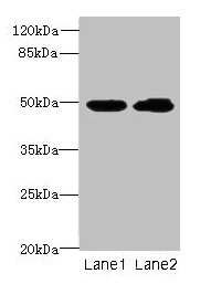
Figure 1. Western blot analysis of HnRNP H/HNRNPH1 using anti-HnRNP H/HNRNPH1 antibody (A07691). Electrophoresis was performed on a 5-20% SDS-PAGE gel at 70V (Stacking gel) / 90V (Resolving gel) for 2-3 hours. The sample well of each lane was loaded with 30 ug of sample under reducing conditions. Lane 1: human 293T whole cell lysates, Lane 2: human Hela whole cell lysates, Lane 3: human HepG2 whole cell lysates, Lane 4: human MCF-7 whole cell lysates, Lane 5: human LNCAP whole cell lysates, Lane 6: human U2OS whole cell lysates, Lane 7: human RT4 whole cell lysates, Lane 8: rat C6 whole cell lysates, Lane 9: rat PC-12 whole cell lysates, Lane 10: mouse NIH/3T3 whole cell lysates. After electrophoresis, proteins were transferred to a nitrocellulose membrane at 150 mA for 50-90 minutes. Blocked the membrane with 5% non-fat milk/TBS for 1.5 hour at RT. The membrane was incubated with rabbit anti-HnRNP H/HNRNPH1 antigen affinity purified polyclonal antibody (Catalog # A07691) at 0.5 microg/mL overnight at 4°C, then washed with TBS-0.1%Tween 3 times with 5 minutes each and probed with a goat anti-rabbit IgG-HRP secondary antibody at a dilution of 1:5000 for 1.5 hour at RT. The signal is developed using an Enhanced Chemiluminescent detection (ECL) kit (Catalog # EK1002) with Tanon 5200 system. A specific band was detected for HnRNP H/HNRNPH1 at approximately 49 kDa. The expected band size for HnRNP H/HNRNPH1 is at 49 kDa.
. HnRNP H/HNRNPH1 was detected in a paraffin-embedded section of human breast cancer tissue. Heat mediated antigen retrieval was performed in EDTA buffer (pH 8.0, epitope retrieval solution). The tissue section was blocked with 10% goat serum. The tissue section was then incubated with 2 microg/ml rabbit anti-HnRNP H/HNRNPH1 Antibody (A07691) overnight at 4°C. Peroxidase Conjugated Goat Anti-rabbit IgG was used as secondary antibody and incubated for 30 minutes at 37°C. The tissue section was developed using HRP Conjugated Rabbit IgG Super Vision Assay Kit (Catalog # SV0002) with DAB as the chromogen.")
. HnRNP H/HNRNPH1 was detected in a paraffin-embedded section of human glioblastoma tissue. Heat mediated antigen retrieval was performed in EDTA buffer (pH 8.0, epitope retrieval solution). The tissue section was blocked with 10% goat serum. The tissue section was then incubated with 2 microg/ml rabbit anti-HnRNP H/HNRNPH1 Antibody (A07691) overnight at 4°C. Peroxidase Conjugated Goat Anti-rabbit IgG was used as secondary antibody and incubated for 30 minutes at 37°C. The tissue section was developed using HRP Conjugated Rabbit IgG Super Vision Assay Kit (Catalog # SV0002) with DAB as the chromogen.")
. HnRNP H/HNRNPH1 was detected in a paraffin-embedded section of human ovarian serous adenocarcinoma tissue. Heat mediated antigen retrieval was performed in EDTA buffer (pH 8.0, epitope retrieval solution). The tissue section was blocked with 10% goat serum. The tissue section was then incubated with 2 microg/ml rabbit anti-HnRNP H/HNRNPH1 Antibody (A07691) overnight at 4°C. Peroxidase Conjugated Goat Anti-rabbit IgG was used as secondary antibody and incubated for 30 minutes at 37°C. The tissue section was developed using HRP Conjugated Rabbit IgG Super Vision Assay Kit (Catalog # SV0002) with DAB as the chromogen.")
. HnRNP H/HNRNPH1 was detected in a paraffin-embedded section of human colorectal adenocarcinoma tissue. Heat mediated antigen retrieval was performed in EDTA buffer (pH 8.0, epitope retrieval solution). The tissue section was blocked with 10% goat serum. The tissue section was then incubated with 2 microg/ml rabbit anti-HnRNP H/HNRNPH1 Antibody (A07691) overnight at 4°C. Peroxidase Conjugated Goat Anti-rabbit IgG was used as secondary antibody and incubated for 30 minutes at 37°C. The tissue section was developed using HRP Conjugated Rabbit IgG Super Vision Assay Kit (Catalog # SV0002) with DAB as the chromogen.")
. HnRNP H/HNRNPH1 was detected in a paraffin-embedded section of human colorectal adenocarcinoma tissue. Heat mediated antigen retrieval was performed in EDTA buffer (pH 8.0, epitope retrieval solution). The tissue section was blocked with 10% goat serum. The tissue section was then incubated with 2 microg/ml rabbit anti-HnRNP H/HNRNPH1 Antibody (A07691) overnight at 4°C. Peroxidase Conjugated Goat Anti-rabbit IgG was used as secondary antibody and incubated for 30 minutes at 37°C. The tissue section was developed using HRP Conjugated Rabbit IgG Super Vision Assay Kit (Catalog # SV0002) with DAB as the chromogen.")
. HnRNP H/HNRNPH1 was detected in a paraffin-embedded section of human tonsil tissue. Heat mediated antigen retrieval was performed in EDTA buffer (pH 8.0, epitope retrieval solution). The tissue section was blocked with 10% goat serum. The tissue section was then incubated with 2 microg/ml rabbit anti-HnRNP H/HNRNPH1 Antibody (A07691) overnight at 4°C. Peroxidase Conjugated Goat Anti-rabbit IgG was used as secondary antibody and incubated for 30 minutes at 37°C. The tissue section was developed using HRP Conjugated Rabbit IgG Super Vision Assay Kit (Catalog # SV0002) with DAB as the chromogen.")
. HnRNP H/HNRNPH1 was detected in a paraffin-embedded section of mouse brain tissue. Heat mediated antigen retrieval was performed in EDTA buffer (pH 8.0, epitope retrieval solution). The tissue section was blocked with 10% goat serum. The tissue section was then incubated with 2 microg/ml rabbit anti-HnRNP H/HNRNPH1 Antibody (A07691) overnight at 4°C. Peroxidase Conjugated Goat Anti-rabbit IgG was used as secondary antibody and incubated for 30 minutes at 37°C. The tissue section was developed using HRP Conjugated Rabbit IgG Super Vision Assay Kit (Catalog # SV0002) with DAB as the chromogen.")
. HnRNP H/HNRNPH1 was detected in a paraffin-embedded section of mouse brain tissue. Heat mediated antigen retrieval was performed in EDTA buffer (pH 8.0, epitope retrieval solution). The tissue section was blocked with 10% goat serum. The tissue section was then incubated with 2 microg/ml rabbit anti-HnRNP H/HNRNPH1 Antibody (A07691) overnight at 4°C. Peroxidase Conjugated Goat Anti-rabbit IgG was used as secondary antibody and incubated for 30 minutes at 37°C. The tissue section was developed using HRP Conjugated Rabbit IgG Super Vision Assay Kit (Catalog # SV0002) with DAB as the chromogen.")
. HnRNP H/HNRNPH1 was detected in a paraffin-embedded section of mouse brain tissue. Heat mediated antigen retrieval was performed in EDTA buffer (pH 8.0, epitope retrieval solution). The tissue section was blocked with 10% goat serum. The tissue section was then incubated with 2 microg/ml rabbit anti-HnRNP H/HNRNPH1 Antibody (A07691) overnight at 4°C. Peroxidase Conjugated Goat Anti-rabbit IgG was used as secondary antibody and incubated for 30 minutes at 37°C. The tissue section was developed using HRP Conjugated Rabbit IgG Super Vision Assay Kit (Catalog # SV0002) with DAB as the chromogen.")
Figure 1. Western blot analysis of HnRNP H/HNRNPH1 using anti-HnRNP H/HNRNPH1 antibody (A07691). Electrophoresis was performed on a 5-20% SDS-PAGE gel at 70V (Stacking gel) / 90V (Resolving gel) for 2-3 hours. The sample well of each lane was loaded with 30 ug of sample under reducing conditions. Lane 1: human 293T whole cell lysates, Lane 2: human Hela whole cell lysates, Lane 3: human HepG2 whole cell lysates, Lane 4: human MCF-7 whole cell lysates, Lane 5: human LNCAP whole cell lysates, Lane 6: human U2OS whole cell lysates, Lane 7: human RT4 whole cell lysates, Lane 8: rat C6 whole cell lysates, Lane 9: rat PC-12 whole cell lysates, Lane 10: mouse NIH/3T3 whole cell lysates. After electrophoresis, proteins were transferred to a nitrocellulose membrane at 150 mA for 50-90 minutes. Blocked the membrane with 5% non-fat milk/TBS for 1.5 hour at RT. The membrane was incubated with rabbit anti-HnRNP H/HNRNPH1 antigen affinity purified polyclonal antibody (Catalog # A07691) at 0.5 microg/mL overnight at 4°C, then washed with TBS-0.1%Tween 3 times with 5 minutes each and probed with a goat anti-rabbit IgG-HRP secondary antibody at a dilution of 1:5000 for 1.5 hour at RT. The signal is developed using an Enhanced Chemiluminescent detection (ECL) kit (Catalog # EK1002) with Tanon 5200 system. A specific band was detected for HnRNP H/HNRNPH1 at approximately 49 kDa. The expected band size for HnRNP H/HNRNPH1 is at 49 kDa.
Anti-HnRNP H/HNRNPH1 Antibody Picoband(r)
A07691-CARRIER-FREE
ApplicationsFlow Cytometry, ImmunoFluorescence, Western Blot, ImmunoCytoChemistry, ImmunoHistoChemistry
Product group Antibodies
ReactivityHuman, Mouse, Rat
TargetHNRNPH1
Overview
- SupplierBoster Bio
- Product NameAnti-HnRNP H/HNRNPH1 Antibody Picoband(r)
- Delivery Days Customer9
- Application Supplier NoteTested Species: In-house tested species with positive results. Other applications have not been tested. Optimal dilutions should be determined by end users.
- ApplicationsFlow Cytometry, ImmunoFluorescence, Western Blot, ImmunoCytoChemistry, ImmunoHistoChemistry
- CertificationResearch Use Only
- ClonalityPolyclonal
- Concentration500 ug/ml
- Gene ID3187
- Target nameHNRNPH1
- Target descriptionheterogeneous nuclear ribonucleoprotein H1
- Target synonymsHNRPH, HNRPH1, NEDCDS, hnRNPH, heterogeneous nuclear ribonucleoprotein H, epididymis secretory sperm binding protein, heterogeneous nuclear ribonucleoprotein H1 (H)
- HostRabbit
- IsotypeIgG
- Protein IDP31943
- Protein NameHeterogeneous nuclear ribonucleoprotein H
- Scientific DescriptionBoster Bio Anti-HnRNP H/HNRNPH1 Antibody Picoband® catalog # A07691. Tested in Flow Cytometry, IF, IHC, ICC, WB applications. This antibody reacts with Human, Mouse, Rat. The brand Picoband indicates this is a premium antibody that guarantees superior quality, high affinity, and strong signals with minimal background in Western blot applications. Only our best-performing antibodies are designated as Picoband, ensuring unmatched performance.
- ReactivityHuman, Mouse, Rat
- Storage Instruction-20°C,2°C to 8°C
- UNSPSC12352203
Related products
Product group Antibodies
HNRNPH1 AntibodyCSB-PA010609HA01HU
ApplicationsWestern Blot, ELISA, ImmunoHistoChemistry
ReactivityHuman
TargetHNRNPH1
- SizePrice
Product group Antibodies
Anti-hnRNP H AntibodyA96577
ApplicationsWestern Blot, ELISA
ReactivityHuman, Mouse, Rat
- SizePrice
Product group Antibodies

HNRNPH1 / hnRNP H AntibodyLS-C334386
ApplicationsWestern Blot
ReactivityHuman, Mouse, Rat
TargetHNRNPH1
- SizePrice
Product group Antibodies
Hnrnph1 Polyclonal AntibodyCAC08523
ApplicationsWestern Blot, ELISA, ImmunoHistoChemistry
TargetHNRNPH1
- SizePrice
Product group Antibodies
HNRNPH1/HNRNPH2 AntibodyPACO03275
ApplicationsWestern Blot, ELISA, ImmunoHistoChemistry
ReactivityHuman, Mouse, Rat
TargetHNRNPH1
- SizePrice
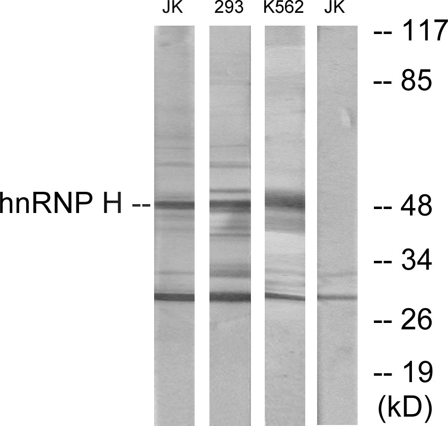
![Non-transfected (–) and transfected (+) 293T whole cell extracts (30 μg) were separated by 10% SDS-PAGE, and the membrane was blotted with hnRNP H antibody [N1C1] (GTX102061) diluted at 1:10000. The HRP-conjugated anti-rabbit IgG antibody (GTX213110-01) was used to detect the primary antibody.](https://www.genetex.com/upload/website/prouct_img/normal/GTX102061/GTX102061_40121_20181005_WB_shRNA_watermark_w_23060100_209.webp)
Product group Antibodies
hnRNP H antibody [N1C1]GTX102061
ApplicationsImmunoFluorescence, Western Blot, ImmunoCytoChemistry, ImmunoHistoChemistry, ImmunoHistoChemistry Paraffin
ReactivityHuman, Mouse
TargetHNRNPH1
- SizePrice
Product group Antibodies
Anti-HNRNPH1 Antibody144-66032
ApplicationsImmunoFluorescence, Western Blot
ReactivityHuman, Mouse, Rat
TargetHNRNPH1
- SizePrice