

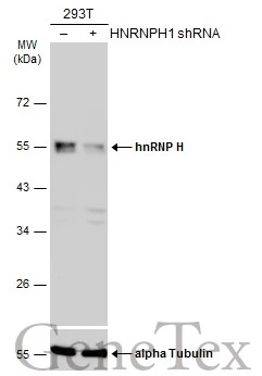
Non-transfected (–) and transfected (+) 293T whole cell extracts (30 μg) were separated by 10% SDS-PAGE, and the membrane was blotted with hnRNP H antibody [N1C1] (GTX102061) diluted at 1:10000. The HRP-conjugated anti-rabbit IgG antibody (GTX213110-01) was used to detect the primary antibody.
![hnRNP H antibody [N1C1] detects hnRNP H protein at nucleus by immunofluorescent analysis. Sample: A431 cells were fixed in 4% paraformaldehyde at RT for 15 min. Green: hnRNP H protein stained by hnRNP H antibody [N1C1] (GTX102061) diluted at 1:500. Red: alpha Tubulin, a cytoskeleton marker, stained by alpha Tubulin antibody [GT114] (GTX628802) diluted at 1:1000. Blue: Hoechst 33342 staining.](https://www.genetex.com/upload/website/prouct_img/normal/GTX102061/GTX102061_40121_20150410_IFA_2_w_23060100_740.webp "hnRNP H antibody [N1C1] detects hnRNP H protein at nucleus by immunofluorescent analysis. Sample: A431 cells were fixed in 4% paraformaldehyde at RT for 15 min. Green: hnRNP H protein stained by hnRNP H antibody [N1C1] (GTX102061) diluted at 1:500. Red: alpha Tubulin, a cytoskeleton marker, stained by alpha Tubulin antibody [GT114] (GTX628802) diluted at 1:1000. Blue: Hoechst 33342 staining.")
 were separated by 10% SDS-PAGE, and the membrane was blotted with hnRNP H antibody (GTX102061) diluted by 1:1000.")
![hnRNP H antibody [N1C1] detects hnRNP H protein at nucleus by immunofluorescent analysis. Sample: HeLa cells were fixed in 4% paraformaldehyde at RT for 15 min. Green: hnRNP H protein stained by hnRNP H antibody [N1C1] (GTX102061) diluted at 1:200. Red: alpha Tubulin, a cytoskeleton marker, stained by alpha Tubulin antibody [B-5-1-2] (GTX11304) diluted at 1:10000. Blue: Hoechst 33342 staining.](https://www.genetex.com/upload/website/prouct_img/normal/GTX102061/GTX102061_40121_20150410_IFA_w_23060100_575.webp "hnRNP H antibody [N1C1] detects hnRNP H protein at nucleus by immunofluorescent analysis. Sample: HeLa cells were fixed in 4% paraformaldehyde at RT for 15 min. Green: hnRNP H protein stained by hnRNP H antibody [N1C1] (GTX102061) diluted at 1:200. Red: alpha Tubulin, a cytoskeleton marker, stained by alpha Tubulin antibody [B-5-1-2] (GTX11304) diluted at 1:10000. Blue: Hoechst 33342 staining.")
 A: Mouse brain 10% SDS PAGE GTX102061 diluted at 1:1000")
 A: A431 (GTX27909) B: H1299 C: Hela D: Hep G2 (GTX27900) 10% SDS PAGE GTX102061 diluted at 1:1000")
 antibody at 1:100 dilution.
Antigen Retrieval: Trilogy? (EDTA based, pH 8.0) buffer, 15min")
Non-transfected (–) and transfected (+) 293T whole cell extracts (30 μg) were separated by 10% SDS-PAGE, and the membrane was blotted with hnRNP H antibody [N1C1] (GTX102061) diluted at 1:10000. The HRP-conjugated anti-rabbit IgG antibody (GTX213110-01) was used to detect the primary antibody.
hnRNP H antibody [N1C1]
GTX102061
ApplicationsImmunoFluorescence, Western Blot, ImmunoCytoChemistry, ImmunoHistoChemistry, ImmunoHistoChemistry Paraffin
Product group Antibodies
ReactivityHuman, Mouse
TargetHNRNPH1
Overview
- SupplierGeneTex
- Product NamehnRNP H antibody [N1C1]
- Delivery Days Customer9
- Application Supplier NoteWB: 1:5000-1:20000. ICC/IF: 1:100-1:1000. IHC-P: 1:100-1:1000. *Optimal dilutions/concentrations should be determined by the researcher.Not tested in other applications.
- ApplicationsImmunoFluorescence, Western Blot, ImmunoCytoChemistry, ImmunoHistoChemistry, ImmunoHistoChemistry Paraffin
- CertificationResearch Use Only
- ClonalityPolyclonal
- Concentration1 mg/ml
- ConjugateUnconjugated
- Gene ID3187
- Target nameHNRNPH1
- Target descriptionheterogeneous nuclear ribonucleoprotein H1
- Target synonymsHNRPH, HNRPH1, NEDCDS, hnRNPH, heterogeneous nuclear ribonucleoprotein H, epididymis secretory sperm binding protein, heterogeneous nuclear ribonucleoprotein H1 (H)
- HostRabbit
- IsotypeIgG
- Protein IDP31943
- Protein NameHeterogeneous nuclear ribonucleoprotein H
- Scientific DescriptionThis gene belongs to the subfamily of ubiquitously expressed heterogeneous nuclear ribonucleoproteins (hnRNPs). The hnRNPs are RNA binding proteins and they complex with heterogeneous nuclear RNA (hnRNA). These proteins are associated with pre-mRNAs in the nucleus and appear to influence pre-mRNA processing and other aspects of mRNA metabolism and transport. While all of the hnRNPs are present in the nucleus, some seem to shuttle between the nucleus and the cytoplasm. The hnRNP proteins have distinct nucleic acid binding properties. The protein encoded by this gene has three repeats of quasi-RRM domains that bind to RNAs. It is very similar to the family member HNRPF. This gene is thought to be potentially involved in hereditary lymphedema type I phenotype. [provided by RefSeq]
- ReactivityHuman, Mouse
- Storage Instruction-20°C or -80°C,2°C to 8°C
- UNSPSC41116161
Datasheet
Related products
Product group Antibodies
HNRNPH1 AntibodyCSB-PA010609HA01HU
ApplicationsWestern Blot, ELISA, ImmunoHistoChemistry
ReactivityHuman
TargetHNRNPH1
- SizePrice
Product group Antibodies
Anti-hnRNP H AntibodyA96577
ApplicationsWestern Blot, ELISA
ReactivityHuman, Mouse, Rat
- SizePrice

Product group Antibodies
Anti-HnRNP H/HNRNPH1 Antibody Picoband(r)A07691-CARRIER-FREE
ApplicationsFlow Cytometry, ImmunoFluorescence, Western Blot, ImmunoCytoChemistry, ImmunoHistoChemistry
ReactivityHuman, Mouse, Rat
TargetHNRNPH1
- SizePrice
Product group Antibodies

HNRNPH1 / hnRNP H AntibodyLS-C334386
ApplicationsWestern Blot
ReactivityHuman, Mouse, Rat
TargetHNRNPH1
- SizePrice
Product group Antibodies
Hnrnph1 Polyclonal AntibodyCAC08523
ApplicationsWestern Blot, ELISA, ImmunoHistoChemistry
TargetHNRNPH1
- SizePrice
Product group Antibodies
HNRNPH1/HNRNPH2 AntibodyPACO03275
ApplicationsWestern Blot, ELISA, ImmunoHistoChemistry
ReactivityHuman, Mouse, Rat
TargetHNRNPH1
- SizePrice
![Non-transfected (–) and transfected (+) 293T whole cell extracts (30 μg) were separated by 10% SDS-PAGE, and the membrane was blotted with hnRNP H antibody [C1C3] (GTX102083) diluted at 1:1000. The HRP-conjugated anti-rabbit IgG antibody (GTX213110-01) was used to detect the primary antibody.](https://www.genetex.com/upload/website/prouct_img/normal/GTX102083/GTX102083_41143_20180907_WB_shRNA_watermark_w_23060100_987.webp)
Product group Antibodies
hnRNP H antibody [C1C3]GTX102083
ApplicationsWestern Blot
ReactivityHuman
TargetHNRNPH1
- SizePrice
Product group Antibodies
Anti-HNRNPH1 Antibody144-66032
ApplicationsImmunoFluorescence, Western Blot
ReactivityHuman, Mouse, Rat
TargetHNRNPH1
- SizePrice