


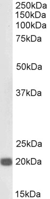
Figure 1. Western blot analysis of HP1 gamma using anti-HP1 gamma antibody (A01142). Electrophoresis was performed on a 5-20% SDS-PAGE gel at 70V (Stacking gel) / 90V (Resolving gel) for 2-3 hours. The sample well of each lane was loaded with 50ug of sample under reducing conditions. Lane 1: rat brain tissue lysates, Lane 2: rat spleen tissue lysate, Lane 3: rat testis tissue lysate, Lane 4: rat PC-12 whole cell lysate, Lane 5: mouse brain tissue lysate, Lane 6: mouse brain tissue lysate, Lane 7: mouse testis tissue lysate, Lane 8: mouse HEPA1-6 whole cell lysate, Lane 9: mouse NIH/3T3 whole cell lysate. After Electrophoresis, proteins were transferred to a Nitrocellulose membrane at 150mA for 50-90 minutes. Blocked the membrane with 5% Non-fat Milk/ TBS for 1.5 hour at RT. The membrane was incubated with rabbit anti-HP1 gamma antigen affinity purified polyclonal antibody (Catalog # A01142) at 0.5 microg/mL overnight at 4°C, then washed with TBS-0.1%Tween 3 times with 5 minutes each and probed with a goat anti-rabbit IgG-HRP secondary antibody at a dilution of 1:10000 for 1.5 hour at RT. The signal is developed using an Enhanced Chemiluminescent detection (ECL) kit (Catalog # EK1002) with Tanon 5200 system. A specific band was detected for HP1 gamma at approximately 21KD. The expected band size for HP1 gamma is at 21KD.
. Electrophoresis was performed on a 5-20% SDS-PAGE gel at 70V (Stacking gel) / 90V (Resolving gel) for 2-3 hours. The sample well of each lane was loaded with 50ug of sample under reducing conditions. Lane 1: human Hela whole cell lysate, Lane 2: human MCF-7 whole cell lysate, Lane 3: human COLO320 whole cell lysate, Lane 4: human HepG2 whole cell lysate, Lane 5: human placenta tissue lysate, Lane 6: human A549 whole cell lysate, Lane 7: human SKOV-3 whole cell lysate, Lane 8: human PANC-1 whole cell lysate. After Electrophoresis, proteins were transferred to a Nitrocellulose membrane at 150mA for 50-90 minutes. Blocked the membrane with 5% Non-fat Milk/ TBS for 1.5 hour at RT. The membrane was incubated with rabbit anti-HP1 gamma antigen affinity purified polyclonal antibody (Catalog # A01142) at 0.5 microg/mL overnight at 4°C, then washed with TBS-0.1%Tween 3 times with 5 minutes each and probed with a goat anti-rabbit IgG-HRP secondary antibody at a dilution of 1:10000 for 1.5 hour at RT. The signal is developed using an Enhanced Chemiluminescent detection (ECL) kit (Catalog # EK1002) with Tanon 5200 system. A specific band was detected for HP1 gamma at approximately 21KD. The expected band size for HP1 gamma is at 21KD.")
. HP1 gamma was detected in paraffin-embedded section of human mammary cancer tissue. Heat mediated antigen retrieval was performed in citrate buffer (pH6, epitope retrieval solution) for 20 mins. The tissue section was blocked with 10% goat serum. The tissue section was then incubated with 1microg/ml rabbit anti-HP1 gamma Antibody (A01142) overnight at 4°C. Biotinylated goat anti-rabbit IgG was used as secondary antibody and incubated for 30 minutes at 37°C. The tissue section was developed using Strepavidin-Biotin-Complex (SABC)(Catalog # SA1022) with DAB as the chromogen.")
. HP1 gamma was detected in paraffin-embedded section of human rectal cancer tissue. Heat mediated antigen retrieval was performed in citrate buffer (pH6, epitope retrieval solution) for 20 mins. The tissue section was blocked with 10% goat serum. The tissue section was then incubated with 1microg/ml rabbit anti-HP1 gamma Antibody (A01142) overnight at 4°C. Biotinylated goat anti-rabbit IgG was used as secondary antibody and incubated for 30 minutes at 37°C. The tissue section was developed using Strepavidin-Biotin-Complex (SABC)(Catalog # SA1022) with DAB as the chromogen.")
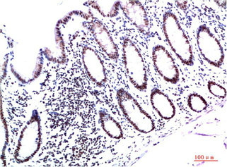
. HP1 gamma was detected in paraffin-embedded section of mouse small intestine tissue. Heat mediated antigen retrieval was performed in citrate buffer (pH6, epitope retrieval solution) for 20 mins. The tissue section was blocked with 10% goat serum. The tissue section was then incubated with 1microg/ml rabbit anti-HP1 gamma Antibody (A01142) overnight at 4°C. Biotinylated goat anti-rabbit IgG was used as secondary antibody and incubated for 30 minutes at 37°C. The tissue section was developed using Strepavidin-Biotin-Complex (SABC)(Catalog # SA1022) with DAB as the chromogen.")
. HP1 gamma was detected in paraffin-embedded section of rat small intestine tissue. Heat mediated antigen retrieval was performed in citrate buffer (pH6, epitope retrieval solution) for 20 mins. The tissue section was blocked with 10% goat serum. The tissue section was then incubated with 1microg/ml rabbit anti-HP1 gamma Antibody (A01142) overnight at 4°C. Biotinylated goat anti-rabbit IgG was used as secondary antibody and incubated for 30 minutes at 37°C. The tissue section was developed using Strepavidin-Biotin-Complex (SABC)(Catalog # SA1022) with DAB as the chromogen.")
. HP1 gamma was detected in immunocytochemical section of U20S cell. Enzyme antigen retrieval was performed using IHC enzyme antigen retrieval reagent (AR0022) for 15 mins. The cells were blocked with 10% goat serum. And then incubated with 2microg/mL rabbit anti-HP1 gamma Antibody (A01142) overnight at 4°C. DyLight®488 Conjugated Goat Anti-Rabbit IgG (BA1127) was used as secondary antibody at 1:100 dilution and incubated for 30 minutes at 37°C. The section was counterstained with DAPI. Visualize using a fluorescence microscope and filter sets appropriate for the label used.")
. Overlay histogram showing A431 cells stained with A01142 (Blue line). To facilitate intracellular staining, cells were fixed with 4% paraformaldehyde and permeabilized with permeabilization buffer. The cells were blocked with 10% normal goat serum. And then incubated with rabbit anti-HP1 gamma Antibody (A01142,1microg/1x106 cells) for 30 min at 20°C. DyLight®488 conjugated goat anti-rabbit IgG (BA1127, 5-10microg/1x106 cells) was used as secondary antibody for 30 minutes at 20°C. Isotype control antibody (Green line) was rabbit IgG (1microg/1x106) used under the same conditions. Unlabelled sample without incubation with primary antibody and secondary antibody (Red line) was used as a blank control.")
. HP1 gamma was detected in frozen section of rat spleen tissues. The tissue section was blocked with 10% goat serum. The tissue section was then incubated with 1microg/ml rabbit anti-HP1 gamma Antibody (A01142) overnight at 4°C. Biotinylated goat anti-rabbit IgG was used as secondary antibody and incubated for 30 minutes at 37°C. The tissue section was developed using Strepavidin-Biotin-Complex (SABC)(Catalog # SA1022) with DAB as the chromogen.")
. HP1 gamma was detected in frozen section of human placenta tissues. The tissue section was blocked with 10% goat serum. The tissue section was then incubated with 1microg/ml rabbit anti-HP1 gamma Antibody (A01142) overnight at 4°C. Biotinylated goat anti-rabbit IgG was used as secondary antibody and incubated for 30 minutes at 37°C. The tissue section was developed using Strepavidin-Biotin-Complex (SABC)(Catalog # SA1022) with DAB as the chromogen.")
Figure 1. Western blot analysis of HP1 gamma using anti-HP1 gamma antibody (A01142). Electrophoresis was performed on a 5-20% SDS-PAGE gel at 70V (Stacking gel) / 90V (Resolving gel) for 2-3 hours. The sample well of each lane was loaded with 50ug of sample under reducing conditions. Lane 1: rat brain tissue lysates, Lane 2: rat spleen tissue lysate, Lane 3: rat testis tissue lysate, Lane 4: rat PC-12 whole cell lysate, Lane 5: mouse brain tissue lysate, Lane 6: mouse brain tissue lysate, Lane 7: mouse testis tissue lysate, Lane 8: mouse HEPA1-6 whole cell lysate, Lane 9: mouse NIH/3T3 whole cell lysate. After Electrophoresis, proteins were transferred to a Nitrocellulose membrane at 150mA for 50-90 minutes. Blocked the membrane with 5% Non-fat Milk/ TBS for 1.5 hour at RT. The membrane was incubated with rabbit anti-HP1 gamma antigen affinity purified polyclonal antibody (Catalog # A01142) at 0.5 microg/mL overnight at 4°C, then washed with TBS-0.1%Tween 3 times with 5 minutes each and probed with a goat anti-rabbit IgG-HRP secondary antibody at a dilution of 1:10000 for 1.5 hour at RT. The signal is developed using an Enhanced Chemiluminescent detection (ECL) kit (Catalog # EK1002) with Tanon 5200 system. A specific band was detected for HP1 gamma at approximately 21KD. The expected band size for HP1 gamma is at 21KD.
Anti-HP1 gamma/CBX3 Antibody Picoband(r)
A01142-CARRIER-FREE
ApplicationsFlow Cytometry, ImmunoFluorescence, Western Blot, ELISA, ImmunoCytoChemistry, ImmunoHistoChemistry, ImmunoHistoChemistry Frozen
Product group Antibodies
ReactivityHuman, Mouse, Rat
TargetCBX3
Overview
- SupplierBoster Bio
- Product NameAnti-HP1 gamma/CBX3 Antibody Picoband(r)
- Delivery Days Customer9
- ApplicationsFlow Cytometry, ImmunoFluorescence, Western Blot, ELISA, ImmunoCytoChemistry, ImmunoHistoChemistry, ImmunoHistoChemistry Frozen
- CertificationResearch Use Only
- ClonalityPolyclonal
- Concentration500 ug/ml
- Gene ID11335
- Target nameCBX3
- Target descriptionchromobox 3
- Target synonymsHECH, HP1-GAMMA, HP1Hs-gamma, HP1gamma, chromobox protein homolog 3, HP1 gamma homolog, chromobox homolog 3 (HP1 gamma homolog, Drosophila), heterochromatin protein 1 homolog gamma, heterochromatin protein HP1 gamma, heterochromatin-like protein 1, modifier 2 protein
- HostRabbit
- IsotypeIgG
- Protein IDQ13185
- Protein NameChromobox protein homolog 3
- Scientific DescriptionBoster Bio Anti-HP1 gamma/CBX3 Antibody Picoband® catalog # A01142. Tested in ELISA, Flow Cytometry, IF, IHC, IHC-F, ICC, WB applications. This antibody reacts with Human, Mouse, Rat. The brand Picoband indicates this is a premium antibody that guarantees superior quality, high affinity, and strong signals with minimal background in Western blot applications. Only our best-performing antibodies are designated as Picoband, ensuring unmatched performance.
- ReactivityHuman, Mouse, Rat
- Storage Instruction-20°C,2°C to 8°C
- UNSPSC12352203
Related products
Product group Antibodies
ReactivityHuman
TargetCBX3
- SizePrice
Product group Antibodies
ApplicationsWestern Blot, ELISA
ReactivityHuman, Mouse
- SizePrice
Product group Antibodies

Anti-CBX3 [RAB-C132]Ab01751-1.1
ApplicationsFlow Cytometry, ImmunoFluorescence, ImmunoPrecipitation
ReactivityHuman
TargetCBX3
- SizePrice
Product group Antibodies

CBX3 / HP1 Gamma Antibody (clone 6A4)LS-C765145
ApplicationsWestern Blot, ImmunoHistoChemistry, ImmunoHistoChemistry Paraffin
ReactivityHuman, Mouse, Rat
TargetCBX3
- SizePrice
Product group Antibodies

ApplicationsWestern Blot, ELISA, ImmunoHistoChemistry
ReactivityCanine, Human, Mouse, Rat
TargetCBX3
- SizePrice

Product group Antibodies
Anti-CBX3 AntibodyHPA004902
ApplicationsWestern Blot, ImmunoCytoChemistry, ImmunoHistoChemistry
ReactivityHuman
TargetCBX3
- SizePrice
Product group Antibodies
CBX3 Monoclonal AntibodyCSB-MA432292
ApplicationsWestern Blot, ELISA, ImmunoHistoChemistry
ReactivityHuman, Mouse, Rat
TargetCBX3
- SizePrice
Product group Antibodies
HP1 gamma Recombinant Antibody, AbBy Fluor-488 ConjugatedBSM-61603R-BF488
ApplicationsFlow Cytometry, ImmunoFluorescence, Western Blot
ReactivityHuman, Mouse, Rat
TargetCBX3
- SizePrice
Product group Antibodies
CBX3 Polyclonal AntibodyCAC13338
ApplicationsImmunoFluorescence, ELISA
TargetCBX3
- SizePrice