


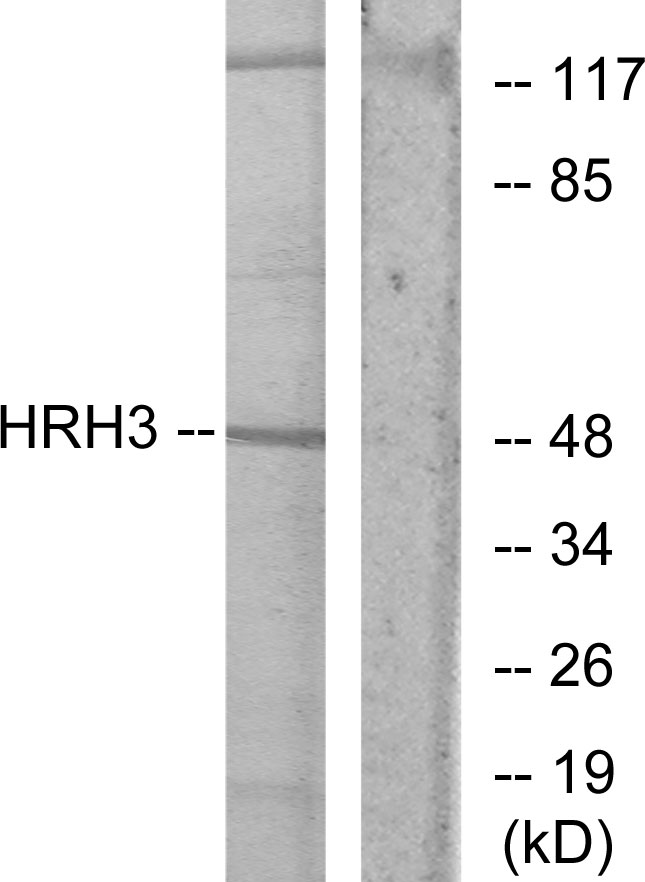
Figure 1. Western blot analysis of HRH3 using anti-HRH3 antibody (M06436). Electrophoresis was performed on a 5-20% SDS-PAGE gel at 70V (Stacking gel) / 90V (Resolving gel) for 2-3 hours. The sample well of each lane was loaded with 30 ug of sample under reducing conditions. Lane 1: human SH-SY5Y whole cell lysates, Lane 2: human MCF-7 whole cell lysates, Lane 3: rat brain tissue lysates, Lane 4: rat C6 whole cell lysates, Lane 5: mouse brain tissue lysates, Lane 6: mouse Neuro-2a whole cell lysates. After electrophoresis, proteins were transferred to a nitrocellulose membrane at 150 mA for 50-90 minutes. Blocked the membrane with 5% non-fat milk/TBS for 1.5 hour at RT. The membrane was incubated with rabbit anti-HRH3 antigen affinity purified monoclonal antibody (M06436) at 1:500 overnight at 4°C, then washed with TBS-0.1%Tween 3 times with 5 minutes each and probed with a goat anti-rabbit IgG-HRP secondary antibody at a dilution of 1:500 for 1.5 hour at RT. The signal is developed using an Enhanced Chemiluminescent detection (ECL) kit (Catalog # EK1002) with Tanon 5200 system. A specific band was detected for HRH3 at approximately 45 kDa. The expected band size for HRH3 is at 49 kDa.
Figure 1. Western blot analysis of HRH3 using anti-HRH3 antibody (M06436). Electrophoresis was performed on a 5-20% SDS-PAGE gel at 70V (Stacking gel) / 90V (Resolving gel) for 2-3 hours. The sample well of each lane was loaded with 30 ug of sample under reducing conditions. Lane 1: human SH-SY5Y whole cell lysates, Lane 2: human MCF-7 whole cell lysates, Lane 3: rat brain tissue lysates, Lane 4: rat C6 whole cell lysates, Lane 5: mouse brain tissue lysates, Lane 6: mouse Neuro-2a whole cell lysates. After electrophoresis, proteins were transferred to a nitrocellulose membrane at 150 mA for 50-90 minutes. Blocked the membrane with 5% non-fat milk/TBS for 1.5 hour at RT. The membrane was incubated with rabbit anti-HRH3 antigen affinity purified monoclonal antibody (M06436) at 1:500 overnight at 4°C, then washed with TBS-0.1%Tween 3 times with 5 minutes each and probed with a goat anti-rabbit IgG-HRP secondary antibody at a dilution of 1:500 for 1.5 hour at RT. The signal is developed using an Enhanced Chemiluminescent detection (ECL) kit (Catalog # EK1002) with Tanon 5200 system. A specific band was detected for HRH3 at approximately 45 kDa. The expected band size for HRH3 is at 49 kDa.
Anti-HRH3 Rabbit Monoclonal Antibody
M06436
ApplicationsFlow Cytometry, ImmunoFluorescence, Western Blot, ImmunoCytoChemistry
Product group Antibodies
ReactivityHuman, Mouse, Rat
TargetHRH3
Overview
- SupplierBoster Bio
- Product NameAnti-HRH3 Rabbit Monoclonal Antibody
- Delivery Days Customer9
- ApplicationsFlow Cytometry, ImmunoFluorescence, Western Blot, ImmunoCytoChemistry
- CertificationResearch Use Only
- ClonalityMonoclonal
- Clone ID24H37
- Gene ID11255
- Target nameHRH3
- Target descriptionhistamine receptor H3
- Target synonymsGPCR97, HH3R, histamine H3 receptor, G protein-coupled receptor 97, H3R
- HostRabbit
- IsotypeIgG
- Protein IDQ9Y5N1
- Protein NameHistamine H3 receptor
- Scientific DescriptionBoster Bio Anti-HRH3 Rabbit Monoclonal Antibody catalog # M06436. Tested in WB, ICC/IF, Flow Cytometry applications. This antibody reacts with Human, Mouse, Rat.
- ReactivityHuman, Mouse, Rat
- Storage Instruction-20°C
- UNSPSC12352203
Related products
Product group Antibodies
Anti-HRH3 AntibodyA98094
ApplicationsWestern Blot, ELISA
ReactivityHuman, Mouse, Rat
- SizePrice
Product group Antibodies
Anti-HRH3 Antibody144-64433
ApplicationsWestern Blot
ReactivityHuman, Rat
TargetHRH3
- SizePrice
Product group Antibodies
References
HRH3 Polyclonal AntibodyBS-3635R
ApplicationsImmunoFluorescence, Western Blot, ImmunoHistoChemistry, ImmunoHistoChemistry Frozen, ImmunoHistoChemistry Paraffin
ReactivityBovine, Canine, Equine, Human, Mouse, Rat
TargetHRH3
- SizePrice
Product group Antibodies
HRH3 AntibodyCSB-PA006801
ApplicationsImmunoFluorescence, Western Blot, ELISA, ImmunoHistoChemistry
ReactivityHuman, Mouse, Rat
TargetHRH3
- SizePrice
Product group Antibodies

Goat anti-HRH3EB09184
ApplicationsELISA
ReactivityBovine, Canine, Human, Mouse, Rat
TargetHRH3
- SizePrice
Product group Antibodies
Hrh3 Polyclonal AntibodyCAC07837
ApplicationsImmunoFluorescence, Western Blot, ELISA, ImmunoHistoChemistry
TargetHRH3
- SizePrice
Product group Antibodies

HRH3 / Histamine 3 Receptor AntibodyLS-C403224
ApplicationsWestern Blot, ELISA, ImmunoHistoChemistry
ReactivityHuman, Mouse
TargetHRH3
- SizePrice
Product group Antibodies
Histamine H3 Receptor antibodyGTX04933
ApplicationsWestern Blot, ImmunoHistoChemistry, ImmunoHistoChemistry Paraffin
ReactivityHuman, Mouse
TargetHRH3
- SizePrice
Product group Antibodies
Anti-HRH3 AntibodyCAB16497
ApplicationsImmunoFluorescence, Western Blot, ELISA, ImmunoCytoChemistry
ReactivityHuman
TargetHRH3
- SizePrice