


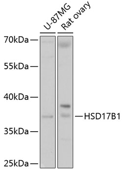
Figure 1. Western blot analysis of HSD17B1 using anti-HSD17B1 antibody (A02198). Electrophoresis was performed on a 5-20% SDS-PAGE gel at 70V (Stacking gel) / 90V (Resolving gel) for 2-3 hours. The sample well of each lane was loaded with 50ug of sample under reducing conditions. Lane 1: Human Placenta tissue lysates. After Electrophoresis, proteins were transferred to a Nitrocellulose membrane at 150mA for 50-90 minutes. Blocked the membrane with 5% Non-fat Milk/ TBS for 1.5 hour at RT. The membrane was incubated with rabbit anti-HSD17B1 antigen affinity purified polyclonal antibody (Catalog # A02198) at 0.5 microg/mL overnight at 4°C, then washed with TBS-0.1%Tween 3 times with 5 minutes each and probed with a goat anti-rabbit IgG-HRP secondary antibody at a dilution of 1:10000 for 1.5 hour at RT. The signal is developed using an Enhanced Chemiluminescent detection (ECL) kit (Catalog # EK1002) with Tanon 5200 system. A specific band was detected for HSD17B1 at approximately 40KD. The expected band size for HSD17B1 is at 35KD.
. HSD17B1 was detected in immunocytochemical section of U20S cells. Enzyme antigen retrieval was performed using IHC enzyme antigen retrieval reagent (AR0022) for 15 mins. The cells were blocked with 10% goat serum. And then incubated with 5microg/mL rabbit anti-HSD17B1 Antibody (A02198) overnight at 4°C. DyLight®488 Conjugated Goat Anti-Rabbit IgG (BA1127) was used as secondary antibody at 1:100 dilution and incubated for 30 minutes at 37°C. The section was counterstained with DAPI. Visualize using a fluorescence microscope and filter sets appropriate for the label used.")
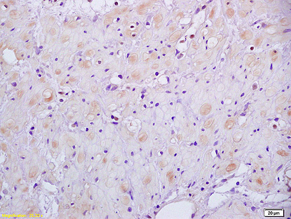
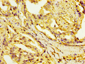
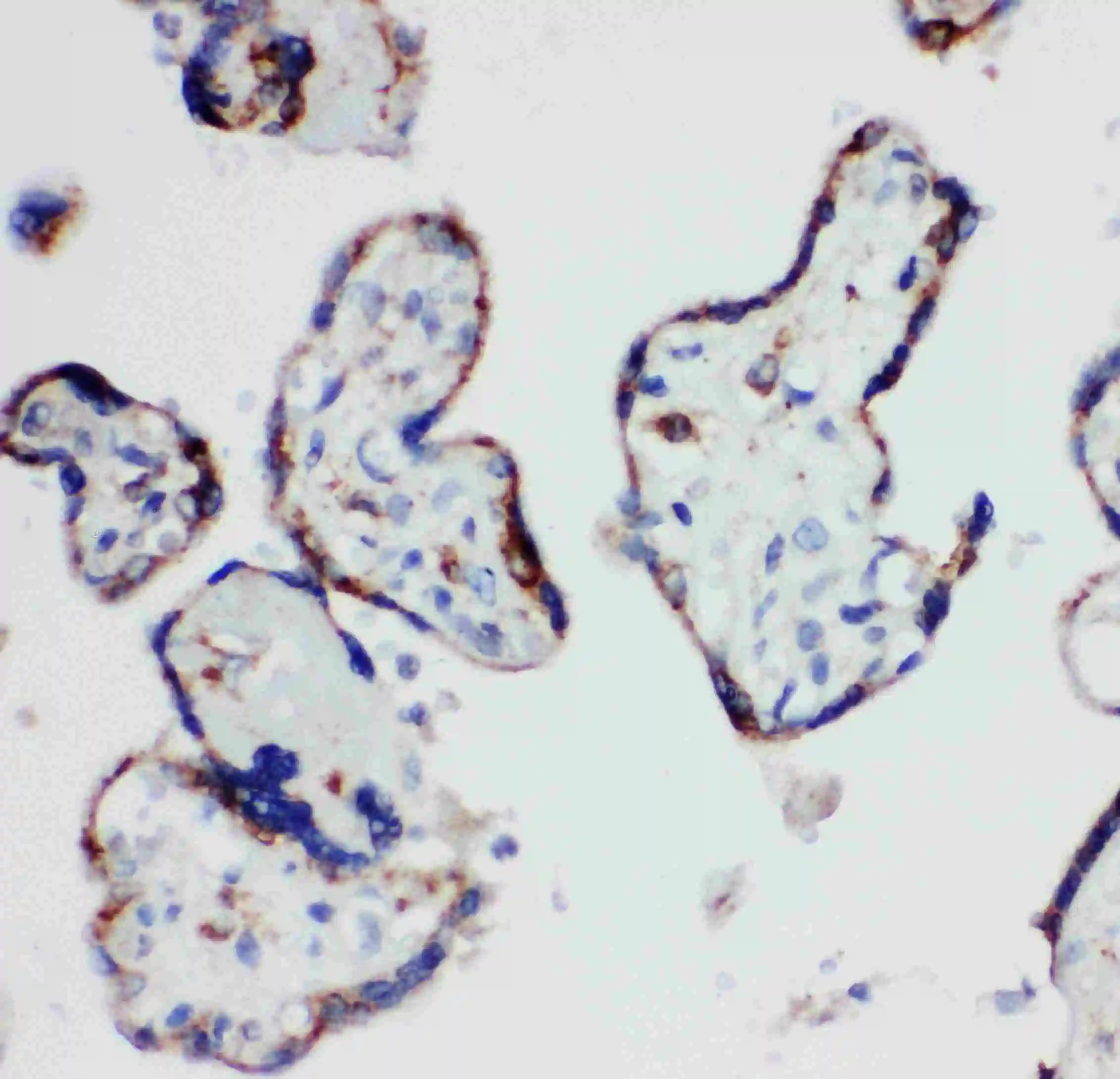
. HSD17B1 was detected in a paraffin-embedded section of human placenta tissue. Heat mediated antigen retrieval was performed in EDTA buffer (pH 8.0, epitope retrieval solution). The tissue section was blocked with 10% goat serum. The tissue section was then incubated with 2 microg/ml rabbit anti-HSD17B1 Antibody (A02198) overnight at 4°C. Peroxidase Conjugated Goat Anti-rabbit IgG was used as secondary antibody and incubated for 30 minutes at 37°C. The tissue section was developed using HRP Conjugated Rabbit IgG Super Vision Assay Kit (Catalog # SV0002) with DAB as the chromogen.")
Figure 1. Western blot analysis of HSD17B1 using anti-HSD17B1 antibody (A02198). Electrophoresis was performed on a 5-20% SDS-PAGE gel at 70V (Stacking gel) / 90V (Resolving gel) for 2-3 hours. The sample well of each lane was loaded with 50ug of sample under reducing conditions. Lane 1: Human Placenta tissue lysates. After Electrophoresis, proteins were transferred to a Nitrocellulose membrane at 150mA for 50-90 minutes. Blocked the membrane with 5% Non-fat Milk/ TBS for 1.5 hour at RT. The membrane was incubated with rabbit anti-HSD17B1 antigen affinity purified polyclonal antibody (Catalog # A02198) at 0.5 microg/mL overnight at 4°C, then washed with TBS-0.1%Tween 3 times with 5 minutes each and probed with a goat anti-rabbit IgG-HRP secondary antibody at a dilution of 1:10000 for 1.5 hour at RT. The signal is developed using an Enhanced Chemiluminescent detection (ECL) kit (Catalog # EK1002) with Tanon 5200 system. A specific band was detected for HSD17B1 at approximately 40KD. The expected band size for HSD17B1 is at 35KD.
Anti-HSD17B1 Antibody Picoband(r)
A02198-CARRIER-FREE
ApplicationsImmunoFluorescence, Western Blot, ELISA, ImmunoCytoChemistry, ImmunoHistoChemistry
Product group Antibodies
ReactivityHuman
TargetHSD17B1
Overview
- SupplierBoster Bio
- Product NameAnti-HSD17B1 Antibody Picoband(r)
- Delivery Days Customer9
- ApplicationsImmunoFluorescence, Western Blot, ELISA, ImmunoCytoChemistry, ImmunoHistoChemistry
- CertificationResearch Use Only
- ClonalityPolyclonal
- Concentration500 ug/ml
- Gene ID3292
- Target nameHSD17B1
- Target descriptionhydroxysteroid 17-beta dehydrogenase 1
- Target synonyms17-beta-HSD, 20-alpha-HSD, E2DH, EDH17B2, EDHB17, HSD17, SDR28C1, 17-beta-hydroxysteroid dehydrogenase type 1, 17-beta-HSD 1, 20 alpha-hydroxysteroid dehydrogenase, estradiol 17-beta-dehydrogenase 1, placental 17-beta-hydroxysteroid dehydrogenase, short chain dehydrogenase/reductase family 28C member 1, short chain dehydrogenase/reductase family 28CE, member 1
- HostRabbit
- IsotypeIgG
- Protein IDP14061
- Protein Name17-beta-hydroxysteroid dehydrogenase type 1
- Scientific DescriptionBoster Bio Anti-HSD17B1 Antibody Picoband® catalog # A02198. Tested in ELISA, IF, IHC, ICC, WB applications. This antibody reacts with Human. The brand Picoband indicates this is a premium antibody that guarantees superior quality, high affinity, and strong signals with minimal background in Western blot applications. Only our best-performing antibodies are designated as Picoband, ensuring unmatched performance.
- ReactivityHuman
- Storage Instruction-20°C,2°C to 8°C
- UNSPSC12352203
Related products
Product group Antibodies
Anti-HSD17B1 AntibodyA16800
ApplicationsImmunoFluorescence, Western Blot, ImmunoCytoChemistry, ImmunoHistoChemistry
ReactivityHuman, Mouse, Rat
- SizePrice
Product group Antibodies
Anti-HSD17B1 Antibody144-61992
ApplicationsImmunoFluorescence, Western Blot, ImmunoHistoChemistry
ReactivityHuman, Mouse, Rat
TargetHSD17B1
- SizePrice
Product group Antibodies
References
HSD17B1 Polyclonal AntibodyBS-3855R
ApplicationsWestern Blot, ELISA, ImmunoHistoChemistry, ImmunoHistoChemistry Paraffin
ReactivityBovine, Canine, Equine, Human, Mouse, Porcine, Rabbit, Rat
TargetHSD17B1
- SizePrice
Product group Antibodies
HSD17B1 AntibodyCSB-PA010766LA01HU
ApplicationsImmunoFluorescence, ELISA, ImmunoHistoChemistry
ReactivityHuman
TargetHSD17B1
- SizePrice
Product group Antibodies
ApplicationsImmunoPrecipitation, Western Blot, ImmunoCytoChemistry, ImmunoHistoChemistry
ReactivityMouse
TargetHSD17B1
- SizePrice
Product group Antibodies

HSD17 / HSD17B1 AntibodyLS-C403226
ApplicationsWestern Blot, ELISA, ImmunoHistoChemistry
ReactivityHuman
TargetHSD17B1
- SizePrice

Product group Antibodies
Anti-HSD17B1 AntibodyHPA021032
ApplicationsImmunoCytoChemistry, ImmunoHistoChemistry
ReactivityHuman
TargetHSD17B1
- SizePrice
Product group Antibodies
HSD17B1 antibodyGTX12312
ApplicationsImmunoFluorescence, Western Blot, ImmunoCytoChemistry, ImmunoHistoChemistry, ImmunoHistoChemistry Paraffin
ReactivityHuman, Mouse, Rat
TargetHSD17B1
- SizePrice