


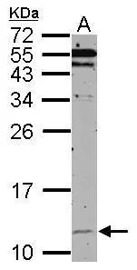
Figure 1. Western blot analysis of HSP10 using anti-HSP10 antibody (M06192-1). Electrophoresis was performed on a 5-20% SDS-PAGE gel at 70V (Stacking gel) / 90V (Resolving gel) for 2-3 hours. The sample well of each lane was loaded with 30 ug of sample under reducing conditions. Lane 1: human A431 whole cell lysates, Lane 2: human A549 whole cell lysates, Lane 3: rat heart tissue lysates, Lane 4: rat skeletal muscle tissue lysates, Lane 5: mouse heart tissue lysates, Lane 6: mouse skeletal muscle tissue lysates. After electrophoresis, proteins were transferred to a nitrocellulose membrane at 150 mA for 50-90 minutes. Blocked the membrane with 5% non-fat milk/TBS for 1.5 hour at RT. The membrane was incubated with rabbit anti-HSP10 antigen affinity purified monoclonal antibody (Catalog # M06192-1) at 1:1000 overnight at 4°C, then washed with TBS-0.1%Tween 3 times with 5 minutes each and probed with a goat anti-rabbit IgG-HRP secondary antibody at a dilution of 1:1000 for 1.5 hour at RT. The signal is developed using an Enhanced Chemiluminescent detection (ECL) kit (Catalog # EK1002) with Tanon 5200 system. A specific band was detected for HSP10 at approximately 11, 13 kDa. The expected band size for HSP10 is at 11 kDa.
. HSP10 was detected in a paraffin-embedded section of human colorectal adenocarcinoma tissue. Heat mediated antigen retrieval was performed in EDTA buffer (pH 8.0, epitope retrieval solution). The tissue section was blocked with 10% goat serum. The tissue section was then incubated with 1:50 rabbit anti-HSP10 Antibody (M06192-1) overnight at 4°C. Peroxidase Conjugated Goat Anti-rabbit IgG was used as secondary antibody and incubated for 30 minutes at 37°C. The tissue section was developed using HRP Conjugated Rabbit IgG Super Vision Assay Kit (Catalog # SV0002) with DAB as the chromogen.")
. HSP10 was detected in a paraffin-embedded section of human lung adenocarcinoma tissue. Heat mediated antigen retrieval was performed in EDTA buffer (pH 8.0, epitope retrieval solution). The tissue section was blocked with 10% goat serum. The tissue section was then incubated with 1:50 rabbit anti-HSP10 Antibody (M06192-1) overnight at 4°C. Peroxidase Conjugated Goat Anti-rabbit IgG was used as secondary antibody and incubated for 30 minutes at 37°C. The tissue section was developed using HRP Conjugated Rabbit IgG Super Vision Assay Kit (Catalog # SV0002) with DAB as the chromogen.")
. HSP10 was detected in a paraffin-embedded section of human prostate cancer tissue. Heat mediated antigen retrieval was performed in EDTA buffer (pH 8.0, epitope retrieval solution). The tissue section was blocked with 10% goat serum. The tissue section was then incubated with 1:50 rabbit anti-HSP10 Antibody (M06192-1) overnight at 4°C. Peroxidase Conjugated Goat Anti-rabbit IgG was used as secondary antibody and incubated for 30 minutes at 37°C. The tissue section was developed using HRP Conjugated Rabbit IgG Super Vision Assay Kit (Catalog # SV0002) with DAB as the chromogen.")
. HSP10 was detected in a paraffin-embedded section of human liver cancer tissue. Heat mediated antigen retrieval was performed in EDTA buffer (pH 8.0, epitope retrieval solution). The tissue section was blocked with 10% goat serum. The tissue section was then incubated with 1:50 rabbit anti-HSP10 Antibody (M06192-1) overnight at 4°C. Peroxidase Conjugated Goat Anti-rabbit IgG was used as secondary antibody and incubated for 30 minutes at 37°C. The tissue section was developed using HRP Conjugated Rabbit IgG Super Vision Assay Kit (Catalog # SV0002) with DAB as the chromogen.")
. HSP10 was detected in a paraffin-embedded section of human spleen tissue. Heat mediated antigen retrieval was performed in EDTA buffer (pH 8.0, epitope retrieval solution). The tissue section was blocked with 10% goat serum. The tissue section was then incubated with 1:50 rabbit anti-HSP10 Antibody (M06192-1) overnight at 4°C. Peroxidase Conjugated Goat Anti-rabbit IgG was used as secondary antibody and incubated for 30 minutes at 37°C. The tissue section was developed using HRP Conjugated Rabbit IgG Super Vision Assay Kit (Catalog # SV0002) with DAB as the chromogen.")
Figure 1. Western blot analysis of HSP10 using anti-HSP10 antibody (M06192-1). Electrophoresis was performed on a 5-20% SDS-PAGE gel at 70V (Stacking gel) / 90V (Resolving gel) for 2-3 hours. The sample well of each lane was loaded with 30 ug of sample under reducing conditions. Lane 1: human A431 whole cell lysates, Lane 2: human A549 whole cell lysates, Lane 3: rat heart tissue lysates, Lane 4: rat skeletal muscle tissue lysates, Lane 5: mouse heart tissue lysates, Lane 6: mouse skeletal muscle tissue lysates. After electrophoresis, proteins were transferred to a nitrocellulose membrane at 150 mA for 50-90 minutes. Blocked the membrane with 5% non-fat milk/TBS for 1.5 hour at RT. The membrane was incubated with rabbit anti-HSP10 antigen affinity purified monoclonal antibody (Catalog # M06192-1) at 1:1000 overnight at 4°C, then washed with TBS-0.1%Tween 3 times with 5 minutes each and probed with a goat anti-rabbit IgG-HRP secondary antibody at a dilution of 1:1000 for 1.5 hour at RT. The signal is developed using an Enhanced Chemiluminescent detection (ECL) kit (Catalog # EK1002) with Tanon 5200 system. A specific band was detected for HSP10 at approximately 11, 13 kDa. The expected band size for HSP10 is at 11 kDa.
Anti-HSP10 Rabbit Monoclonal Antibody
M06192-1
ApplicationsImmunoFluorescence, ImmunoPrecipitation, Western Blot, ImmunoCytoChemistry, ImmunoHistoChemistry
Product group Antibodies
ReactivityHuman, Mouse, Rat
TargetHSPE1
Overview
- SupplierBoster Bio
- Product NameAnti-HSP10 Rabbit Monoclonal Antibody
- Delivery Days Customer9
- ApplicationsImmunoFluorescence, ImmunoPrecipitation, Western Blot, ImmunoCytoChemistry, ImmunoHistoChemistry
- CertificationResearch Use Only
- ClonalityMonoclonal
- Clone ID19H75
- Gene ID3336
- Target nameHSPE1
- Target descriptionheat shock protein family E (Hsp10) member 1
- Target synonymsCPN10, EPF, GROES, HSP10, 10 kDa heat shock protein, mitochondrial, 10 kDa chaperonin, chaperonin 10, early-pregnancy factor, epididymis secretory sperm binding protein, heat shock 10kD protein 1 (chaperonin 10), heat shock 10kDa protein 1, heat shock protein family E member 1
- HostRabbit
- IsotypeIgG
- Protein IDP61604
- Protein Name10 kDa heat shock protein, mitochondrial
- Scientific DescriptionBoster Bio Anti-HSP10 Rabbit Monoclonal Antibody catalog # M06192-1. Tested in WB, IHC, ICC/IF, IP applications. This antibody reacts with Human, Mouse, Rat.
- ReactivityHuman, Mouse, Rat
- Storage Instruction-20°C
- UNSPSC12352203
Related products

Product group Antibodies
HSPE1 AntibodyCSB-PA009331
ApplicationsImmunoFluorescence, Western Blot, ELISA, ImmunoHistoChemistry
ReactivityHuman, Mouse, Rat
TargetHSPE1
- SizePrice
Product group Antibodies
ReactivityHuman
TargetHSPE1
- SizePrice
Product group Antibodies
Anti-HSP10 AntibodyA95222
ApplicationsImmunoFluorescence, Western Blot, ELISA, ImmunoHistoChemistry
ReactivityHuman, Mouse, Rat
- SizePrice

Product group Antibodies
Anti-HSPE1 AntibodyHPA038755
ApplicationsWestern Blot, ImmunoHistoChemistry
ReactivityHuman
TargetHSPE1
- SizePrice
Product group Antibodies

Goat anti-CPN10 / HSPE1EB12835
ApplicationsWestern Blot, ELISA, ImmunoHistoChemistry
ReactivityBovine, Canine, Human, Mouse, Porcine, Rat
TargetHSPE1
- SizePrice
Product group Antibodies

HSPE1 / HSP10 / Chaperonin 10 AntibodyLS-C343047
ApplicationsWestern Blot, ELISA
ReactivityHuman
TargetHSPE1
- SizePrice
Product group Antibodies
Hspe1 Polyclonal AntibodyCAC10410
ApplicationsELISA, ImmunoHistoChemistry
TargetHSPE1
- SizePrice
Product group Antibodies
HSPE1 Recombinant AntibodyBSM-60471R
ApplicationsImmunoFluorescence, Western Blot, ImmunoCytoChemistry, ImmunoHistoChemistry, ImmunoHistoChemistry Frozen, ImmunoHistoChemistry Paraffin
ReactivityHuman, Mouse, Rat
TargetHSPE1
- SizePrice
Product group Antibodies
Cpn10 antibodyGTX102296
ApplicationsWestern Blot
ReactivityHuman, Mouse
TargetHSPE1
- SizePrice
Product group Antibodies
Anti-HSP10 Antibody130-10221
ApplicationsWestern Blot, ELISA
TargetHSPE1
- SizePrice