


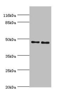
Figure 1. Western blot analysis of Hsp47/SERPINH1 using anti-Hsp47/SERPINH1 antibody (PB9325). Electrophoresis was performed on a 5-20% SDS-PAGE gel at 70V (Stacking gel) / 90V (Resolving gel) for 2-3 hours. The sample well of each lane was loaded with 30 ug of sample under reducing conditions. Lane 1: human A431 whole cell lysates, Lane 2: human SK-N-SH whole cell lysates, Lane 3: human Hela whole cell lysates, Lane 4: rat lung tissue lysates, Lane 5: rat kidney tissue lysates, Lane 6: rat L6 whole cell lysates, Lane 7: mouse lung tissue lysates, Lane 8: mouse NIH/3T3 whole cell lysates. After electrophoresis, proteins were transferred to a nitrocellulose membrane at 150 mA for 50-90 minutes. Blocked the membrane with 5% non-fat milk/TBS for 1.5 hour at RT. The membrane was incubated with rabbit anti-Hsp47/SERPINH1 antigen affinity purified polyclonal antibody (Catalog # PB9325) at 0.5 microg/mL overnight at 4°C, then washed with TBS-0.1%Tween 3 times with 5 minutes each and probed with a goat anti-rabbit IgG-HRP secondary antibody at a dilution of 1:5000 for 1.5 hour at RT. The signal is developed using an Enhanced Chemiluminescent detection (ECL) kit (Catalog # EK1002) with Tanon 5200 system. A specific band was detected for Hsp47/SERPINH1 at approximately 46 kDa. The expected band size for Hsp47/SERPINH1 is at 46 kDa.
. Hsp47/SERPINH1 was detected in a paraffin-embedded section of human breast cancer tissue. Heat mediated antigen retrieval was performed in EDTA buffer (pH 8.0, epitope retrieval solution). The tissue section was blocked with 10% goat serum. The tissue section was then incubated with 2 microg/ml rabbit anti-Hsp47/SERPINH1 Antibody (PB9325) overnight at 4°C. Peroxidase Conjugated Goat Anti-rabbit IgG was used as secondary antibody and incubated for 30 minutes at 37°C. The tissue section was developed using HRP Conjugated Rabbit IgG Super Vision Assay Kit (Catalog # SV0002) with DAB as the chromogen.")
. Hsp47/SERPINH1 was detected in a paraffin-embedded section of human endometrial adenocarcinoma tissue. Heat mediated antigen retrieval was performed in EDTA buffer (pH 8.0, epitope retrieval solution). The tissue section was blocked with 10% goat serum. The tissue section was then incubated with 2 microg/ml rabbit anti-Hsp47/SERPINH1 Antibody (PB9325) overnight at 4°C. Peroxidase Conjugated Goat Anti-rabbit IgG was used as secondary antibody and incubated for 30 minutes at 37°C. The tissue section was developed using HRP Conjugated Rabbit IgG Super Vision Assay Kit (Catalog # SV0002) with DAB as the chromogen.")
. Hsp47/SERPINH1 was detected in a paraffin-embedded section of human esophageal squamous carcinoma tissue. Heat mediated antigen retrieval was performed in EDTA buffer (pH 8.0, epitope retrieval solution). The tissue section was blocked with 10% goat serum. The tissue section was then incubated with 2 microg/ml rabbit anti-Hsp47/SERPINH1 Antibody (PB9325) overnight at 4°C. Peroxidase Conjugated Goat Anti-rabbit IgG was used as secondary antibody and incubated for 30 minutes at 37°C. The tissue section was developed using HRP Conjugated Rabbit IgG Super Vision Assay Kit (Catalog # SV0002) with DAB as the chromogen.")
. Hsp47/SERPINH1 was detected in a paraffin-embedded section of human liver cancer tissue. Heat mediated antigen retrieval was performed in EDTA buffer (pH 8.0, epitope retrieval solution). The tissue section was blocked with 10% goat serum. The tissue section was then incubated with 2 microg/ml rabbit anti-Hsp47/SERPINH1 Antibody (PB9325) overnight at 4°C. Peroxidase Conjugated Goat Anti-rabbit IgG was used as secondary antibody and incubated for 30 minutes at 37°C. The tissue section was developed using HRP Conjugated Rabbit IgG Super Vision Assay Kit (Catalog # SV0002) with DAB as the chromogen.")
. Hsp47/SERPINH1 was detected in a paraffin-embedded section of human spleen tissue. Heat mediated antigen retrieval was performed in EDTA buffer (pH 8.0, epitope retrieval solution). The tissue section was blocked with 10% goat serum. The tissue section was then incubated with 2 microg/ml rabbit anti-Hsp47/SERPINH1 Antibody (PB9325) overnight at 4°C. Peroxidase Conjugated Goat Anti-rabbit IgG was used as secondary antibody and incubated for 30 minutes at 37°C. The tissue section was developed using HRP Conjugated Rabbit IgG Super Vision Assay Kit (Catalog # SV0002) with DAB as the chromogen.")
. Hsp47/SERPINH1 was detected in a paraffin-embedded section of human lung adenocarcinoma tissue. Heat mediated antigen retrieval was performed in EDTA buffer (pH 8.0, epitope retrieval solution). The tissue section was blocked with 10% goat serum. The tissue section was then incubated with 2 microg/ml rabbit anti-Hsp47/SERPINH1 Antibody (PB9325) overnight at 4°C. Peroxidase Conjugated Goat Anti-rabbit IgG was used as secondary antibody and incubated for 30 minutes at 37°C. The tissue section was developed using HRP Conjugated Rabbit IgG Super Vision Assay Kit (Catalog # SV0002) with DAB as the chromogen.")
Figure 1. Western blot analysis of Hsp47/SERPINH1 using anti-Hsp47/SERPINH1 antibody (PB9325). Electrophoresis was performed on a 5-20% SDS-PAGE gel at 70V (Stacking gel) / 90V (Resolving gel) for 2-3 hours. The sample well of each lane was loaded with 30 ug of sample under reducing conditions. Lane 1: human A431 whole cell lysates, Lane 2: human SK-N-SH whole cell lysates, Lane 3: human Hela whole cell lysates, Lane 4: rat lung tissue lysates, Lane 5: rat kidney tissue lysates, Lane 6: rat L6 whole cell lysates, Lane 7: mouse lung tissue lysates, Lane 8: mouse NIH/3T3 whole cell lysates. After electrophoresis, proteins were transferred to a nitrocellulose membrane at 150 mA for 50-90 minutes. Blocked the membrane with 5% non-fat milk/TBS for 1.5 hour at RT. The membrane was incubated with rabbit anti-Hsp47/SERPINH1 antigen affinity purified polyclonal antibody (Catalog # PB9325) at 0.5 microg/mL overnight at 4°C, then washed with TBS-0.1%Tween 3 times with 5 minutes each and probed with a goat anti-rabbit IgG-HRP secondary antibody at a dilution of 1:5000 for 1.5 hour at RT. The signal is developed using an Enhanced Chemiluminescent detection (ECL) kit (Catalog # EK1002) with Tanon 5200 system. A specific band was detected for Hsp47/SERPINH1 at approximately 46 kDa. The expected band size for Hsp47/SERPINH1 is at 46 kDa.
Anti-Hsp47/SERPINH1 Antibody Picoband(r)
PB9325-CARRIER-FREE
ApplicationsWestern Blot, ImmunoHistoChemistry
Product group Antibodies
ReactivityHuman, Mouse, Rat
TargetSERPINH1
Overview
- SupplierBoster Bio
- Product NameAnti-Hsp47/SERPINH1 Antibody Picoband(r)
- Delivery Days Customer9
- Application Supplier NoteWB: The detection limit for Hsp47 is approximately 0.25ng/lane under reducing conditions. Tested Species: In-house tested species with positive results. By Heat: Boiling the paraffin sections in 10mM citrate buffer, pH6.0, for 20mins is required for the staining of formalin/paraffin sections. Other applications have not been tested. Optimal dilutions should be determined by end users.
- ApplicationsWestern Blot, ImmunoHistoChemistry
- CertificationResearch Use Only
- ClonalityPolyclonal
- Concentration500 ug/ml
- Gene ID871
- Target nameSERPINH1
- Target descriptionserpin family H member 1
- Target synonymsAsTP3, CBP1, CBP2, HSP47, OI10, PIG14, PPROM, RA-A47, SERPINH2, gp46, serpin H1, 47 kDa heat shock protein, arsenic-transactivated protein 3, cell proliferation-inducing gene 14 protein, collagen binding protein 1, colligin-1, colligin-2, heat shock protein 47, rheumatoid arthritis antigen A-47, rheumatoid arthritis-related antigen RA-A47, serine (or cysteine) proteinase inhibitor, clade H (heat shock protein 47), member 1, (collagen binding protein 1), serine (or cysteine) proteinase inhibitor, clade H (heat shock protein 47), member 2, (collagen-binding protein 2), serpin peptidase inhibitor, clade H (heat shock protein 47), member 1, (collagen binding protein 1)
- HostRabbit
- IsotypeIgG
- Protein IDP50454
- Protein NameSerpin H1
- Scientific DescriptionBoster Bio Anti-Hsp47/SERPINH1 Antibody Picoband® catalog # PB9325. Tested in IHC, WB applications. This antibody reacts with Human, Mouse, Rat. The brand Picoband indicates this is a premium antibody that guarantees superior quality, high affinity, and strong signals with minimal background in Western blot applications. Only our best-performing antibodies are designated as Picoband, ensuring unmatched performance.
- ReactivityHuman, Mouse, Rat
- Storage Instruction-20°C,2°C to 8°C
- UNSPSC12352203
Related products
Product group Antibodies
ReactivityHuman
TargetSERPINH1
- SizePrice
Product group Antibodies
SERPINH1 AntibodyCSB-PA021087ESR2HU
ApplicationsWestern Blot, ELISA, ImmunoHistoChemistry
ReactivityHuman
TargetSERPINH1
- SizePrice

Product group Antibodies
Anti-SERPINH1 AntibodyA34813
ApplicationsImmunoFluorescence, Western Blot, ImmunoHistoChemistry
ReactivityHuman, Mouse, Rat
- SizePrice
Product group Antibodies

SERPINH1 / HSP47 AntibodyLS-C831104
ApplicationsWestern Blot, ELISA
ReactivityHuman, Mouse, Rat
TargetSERPINH1
- SizePrice

Product group Antibodies
Anti-SERPINH1 AntibodyHPA029198
ApplicationsWestern Blot, ImmunoCytoChemistry, ImmunoHistoChemistry
ReactivityHuman, Mouse
TargetSERPINH1
- SizePrice
Product group Antibodies
Serpinh1 Polyclonal AntibodyCAC10563
ApplicationsWestern Blot, ELISA, ImmunoHistoChemistry
TargetSERPINH1
- SizePrice
Product group Antibodies
HSP47 Polyclonal AntibodyBS-1538R
ApplicationsImmunoFluorescence, ELISA, ImmunoHistoChemistry, ImmunoHistoChemistry Frozen, ImmunoHistoChemistry Paraffin
ReactivityHuman, Mouse, Rat
TargetSERPINH1
- SizePrice
![HSP47 antibody [N2C2], Internal detects HSP47 protein at cytosol on human breast carcinoma by immunohistochemical analysis. Sample: Paraffin-embedded human breast carcinoma. HSP47 antibody [N2C2], Internal (GTX103011) dilution: 1:500.
Antigen Retrieval: Trilogy? (EDTA based, pH 8.0) buffer, 15min](https://www.genetex.com/upload/website/prouct_img/normal/GTX103011/GTX103011_40150_IHC_w_23060119_320.webp)
Product group Antibodies
HSP47 antibody [N2C2], InternalGTX103011
ApplicationsWestern Blot, ImmunoHistoChemistry, ImmunoHistoChemistry Paraffin
ReactivityHuman, Mouse
TargetSERPINH1
- SizePrice
Product group Antibodies
Anti-Rat Serpinh1 (Center) Antibody102-20009
ApplicationsWestern Blot, ImmunoHistoChemistry, ImmunoHistoChemistry Paraffin
TargetSERPINH1
- SizePrice