

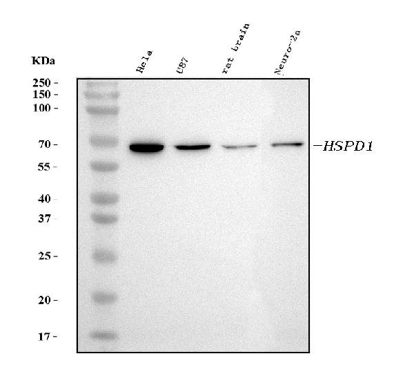
Figure 1. Western blot analysis of HSP60 using anti-HSP60 antibody (MA1049). Electrophoresis was performed on a 5-20% SDS-PAGE gel at 70V (Stacking gel) / 90V (Resolving gel) for 2-3 hours. The sample well of each lane was loaded with 30 ug of sample under reducing conditions. Lane 1: human Hela whole cell lysates, Lane 2: human U87 whole cell lysates, Lane 3: rat brain tissue lysates, Lane 4: mouse Neuro-2a whole cell lysates. After electrophoresis, proteins were transferred to a nitrocellulose membrane at 150 mA for 50-90 minutes. Blocked the membrane with 5% non-fat milk/TBS for 1.5 hour at RT. The membrane was incubated with mouse anti-HSP60 antigen affinity purified monoclonal antibody (Catalog # MA1049) at 2 microg/mL overnight at 4°C, then washed with TBS-0.1%Tween 3 times with 5 minutes each and probed with a goat anti-mouse IgG-HRP secondary antibody at a dilution of 1:5000 for 1.5 hour at RT. The signal is developed using an Enhanced Chemiluminescent detection (ECL) kit (Catalog # EK1001) with Tanon 5200 system. A specific band was detected for HSP60 at approximately 65 kDa. The expected band size for HSP60 is at 65 kDa.
Figure 1. Western blot analysis of HSP60 using anti-HSP60 antibody (MA1049). Electrophoresis was performed on a 5-20% SDS-PAGE gel at 70V (Stacking gel) / 90V (Resolving gel) for 2-3 hours. The sample well of each lane was loaded with 30 ug of sample under reducing conditions. Lane 1: human Hela whole cell lysates, Lane 2: human U87 whole cell lysates, Lane 3: rat brain tissue lysates, Lane 4: mouse Neuro-2a whole cell lysates. After electrophoresis, proteins were transferred to a nitrocellulose membrane at 150 mA for 50-90 minutes. Blocked the membrane with 5% non-fat milk/TBS for 1.5 hour at RT. The membrane was incubated with mouse anti-HSP60 antigen affinity purified monoclonal antibody (Catalog # MA1049) at 2 microg/mL overnight at 4°C, then washed with TBS-0.1%Tween 3 times with 5 minutes each and probed with a goat anti-mouse IgG-HRP secondary antibody at a dilution of 1:5000 for 1.5 hour at RT. The signal is developed using an Enhanced Chemiluminescent detection (ECL) kit (Catalog # EK1001) with Tanon 5200 system. A specific band was detected for HSP60 at approximately 65 kDa. The expected band size for HSP60 is at 65 kDa.
Anti-HSP60 Hspd1 Antibody (Monoclonal, LK1)
MA1049
ApplicationsWestern Blot, ImmunoHistoChemistry
Product group Antibodies
TargetHSPD1
Overview
- SupplierBoster Bio
- Product NameAnti-HSP60 Antibody (Monoclonal, LK1)
- Delivery Days Customer9
- Application Supplier NoteOther applications have not been tested. Optimal dilutions should be determined by end users.
- ApplicationsWestern Blot, ImmunoHistoChemistry
- Applications SupplierIHP, WB, IHC
- CertificationResearch Use Only
- ClonalityMonoclonal
- Clone IDLK1
- Concentration100 ug/ml
- Gene ID3329
- Target nameHSPD1
- Target descriptionheat shock protein family D (Hsp60) member 1
- Target synonymsCPN60, GROEL, HLD4, HSP-60, HSP60, HSP65, HuCHA60, SPG13, 60 kDa heat shock protein, mitochondrial, 60 kDa chaperonin, P60 lymphocyte protein, chaperonin 60, epididymis secretory sperm binding protein, heat shock 60kDa protein 1 (chaperonin), heat shock protein 65, heat shock protein family D member 1, mitochondrial matrix protein P1, short heat shock protein 60 Hsp60s1
- HostMouse
- IsotypeIgG1
- Protein IDP63039
- Protein Name60 kDa heat shock protein, mitochondrial
- Scientific DescriptionBoster Bio Anti-HSP60 Hspd1 Antibody (Monoclonal, LK1) catalog # MA1049. Tested in IHC, WB applications. This antibody reacts with Chicken, Human, Mouse, Rat.
- Reactivity SupplierChicken, Human, Rat, Monkey
- Storage Instruction-20°C,2°C to 8°C
- UNSPSC12352203
References
- Dündar AS, Oruç M, Celbiş O, et al. An experimental rat model of electric shock injury with isolated electric shock and water conduction: the histopathological changes on the skin and internal organs and the effect on biochemical parameters. Int J Legal Med. 2023,137(1):215-226. doi: 10.1007/s00414-022-02834-wRead this paper
- Li L, Nan P, Zhai S, et al. Molecular cloning, characterization, and expression of hsp60 in caudal fin regeneration of Misgurnus anguillicaudatus. Mol Cell Biochem. 2014,387(1-2):143-50. doi: 10.1007/s11010-013-1879-0Read this paper
Datasheet
MSDS
Related products
Product group Antibodies
ApplicationsFlow Cytometry
TargetHSPD1
- SizePrice
Product group Antibodies
Anti-Hsp60 AntibodyA83554
ApplicationsImmunoFluorescence, Western Blot, ELISA, ImmunoHistoChemistry
- SizePrice
Product group Antibodies
References
HSP60 Polyclonal AntibodyBS-0191R
ApplicationsImmunoFluorescence, Western Blot, ELISA, ImmunoCytoChemistry, ImmunoHistoChemistry, ImmunoHistoChemistry Frozen, ImmunoHistoChemistry Paraffin
TargetHSPD1
- SizePrice
Product group Antibodies

ApplicationsImmunoFluorescence, Western Blot, ELISA, ImmunoHistoChemistry
TargetHSPD1
- SizePrice
Product group Antibodies
References
HSP60 antibodyGTX110089
ApplicationsImmunoFluorescence, Western Blot, ELISA, ImmunoCytoChemistry, ImmunoHistoChemistry, ImmunoHistoChemistry Paraffin
TargetHSPD1
- SizePrice
Product group Antibodies

ApplicationsELISA
TargetHSPD1
- SizePrice

Product group Antibodies
Anti-HSPD1 AntibodyHPA001523
ApplicationsWestern Blot, ImmunoCytoChemistry, ImmunoHistoChemistry
ReactivityHuman, Mouse, Rat
TargetHSPD1
- SizePrice