


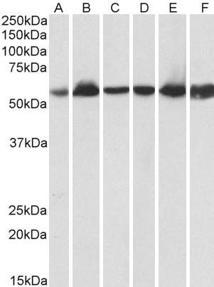
Figure 1. Western blot analysis of HSPD1 using anti-HSPD1 antibody (M01280-3). Electrophoresis was performed on a 5-20% SDS-PAGE gel at 70V (Stacking gel) / 90V (Resolving gel) for 2-3 hours. The sample well of each lane was loaded with 50ug of sample under reducing conditions. Lane 1: human Caco-2 whole cell lysates Lane 2: human A549 whole cell lysates Lane 3: human THP-1 whole cell lysates Lane 4: human SW620 whole cell lysates Lane 5: human U-937 whole cell lysates Lane 6: human HepG2 whole cell lysates Lane 7: rat RH35 whole cell lysates Lane 8: mouse RAW246.7 whole cell lysates After Electrophoresis, proteins were transferred to a Nitrocellulose membrane at 150mA for 50-90 minutes. Blocked the membrane with 5% Non-fat Milk/ TBS for 1.5 hour at RT. The membrane was incubated with mouse anti-HSPD1 antigen affinity purified monoclonal antibody (Catalog # M01280-3) at 0.5 microg/mL overnight at 4°C, then washed with TBS-0.1%Tween 3 times with 5 minutes each and probed with a goat anti-mouse IgG-HRP secondary antibody at a dilution of 1:10000 for 1.5 hour at RT. The signal is developed using an Enhanced Chemiluminescent detection (ECL) kit (Catalog # EK1001) with Tanon 5200 system. A specific band was detected for HSPD1 at approximately 60KD. The expected band size for HSPD1 is at 60KD.
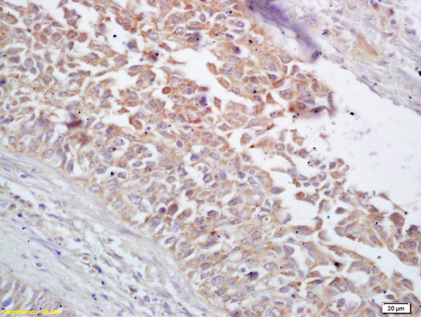
. HSPD1 was detected in paraffin-embedded section of human intestinal cancer tissues. Heat mediated antigen retrieval was performed in citrate buffer (pH6, epitope retrieval solution) for 20 mins. The tissue section was blocked with 10% goat serum. The tissue section was then incubated with 1microg/ml mouse anti-HSPD1 Antibody (M01280-3) overnight at 4°C. Biotinylated goat anti-mouse IgG was used as secondary antibody and incubated for 30 minutes at 37°C. The tissue section was developed using Strepavidin-Biotin-Complex (SABC)(Catalog # SA1021) with DAB as the chromogen.")
. HSPD1 was detected in paraffin-embedded section of human lung cancer tissues. Heat mediated antigen retrieval was performed in citrate buffer (pH6, epitope retrieval solution) for 20 mins. The tissue section was blocked with 10% goat serum. The tissue section was then incubated with 1microg/ml mouse anti-HSPD1 Antibody (M01280-3) overnight at 4°C. Biotinylated goat anti-mouse IgG was used as secondary antibody and incubated for 30 minutes at 37°C. The tissue section was developed using Strepavidin-Biotin-Complex (SABC)(Catalog # SA1021) with DAB as the chromogen.")
. HSPD1 was detected in paraffin-embedded section of human mammary cancer tissues. Heat mediated antigen retrieval was performed in citrate buffer (pH6, epitope retrieval solution) for 20 mins. The tissue section was blocked with 10% goat serum. The tissue section was then incubated with 1microg/ml mouse anti-HSPD1 Antibody (M01280-3) overnight at 4°C. Biotinylated goat anti-mouse IgG was used as secondary antibody and incubated for 30 minutes at 37°C. The tissue section was developed using Strepavidin-Biotin-Complex (SABC)(Catalog # SA1021) with DAB as the chromogen.")
. HSPD1 was detected in paraffin-embedded section of mouse liver tissues. Heat mediated antigen retrieval was performed in citrate buffer (pH6, epitope retrieval solution) for 20 mins. The tissue section was blocked with 10% goat serum. The tissue section was then incubated with 1microg/ml mouse anti-HSPD1 Antibody (M01280-3) overnight at 4°C. Biotinylated goat anti-mouse IgG was used as secondary antibody and incubated for 30 minutes at 37°C. The tissue section was developed using Strepavidin-Biotin-Complex (SABC)(Catalog # SA1021) with DAB as the chromogen.")
. HSPD1 was detected in paraffin-embedded section of rat liver tissues. Heat mediated antigen retrieval was performed in citrate buffer (pH6, epitope retrieval solution) for 20 mins. The tissue section was blocked with 10% goat serum. The tissue section was then incubated with 1microg/ml mouse anti-HSPD1 Antibody (M01280-3) overnight at 4°C. Biotinylated goat anti-mouse IgG was used as secondary antibody and incubated for 30 minutes at 37°C. The tissue section was developed using Strepavidin-Biotin-Complex (SABC)(Catalog # SA1021) with DAB as the chromogen.")
. Overlay histogram showing A431 cells stained with M01280-3 (Blue line). To facilitate intracellular staining, cells were fixed with 4% paraformaldehyde and permeabilized with permeabilization buffer. The cells were blocked with 10% normal goat serum. And then incubated with mouse anti-HSPD1 Antibody (M01280-3,1microg/1x106 cells) for 30 min at 20°C. DyLight®488 conjugated goat anti-mouse IgG (BA1126, 5-10microg/1x106 cells) was used as secondary antibody for 30 minutes at 20°C. Isotype control antibody (Green line) was mouse IgG (1microg/1x106) used under the same conditions. Unlabelled sample without incubation with primary antibody and secondary antibody (Red line) was used as a blank control.")
. Overlay histogram showing HepG2 cells stained with M01280-3 (Blue line). To facilitate intracellular staining, cells were fixed with 4% paraformaldehyde and permeabilized with permeabilization buffer. The cells were blocked with 10% normal goat serum. And then incubated with mouse anti-HSPD1 Antibody (M01280-3,1microg/1x106 cells) for 30 min at 20°C. DyLight®488 conjugated goat anti-mouse IgG (BA1126, 5-10microg/1x106 cells) was used as secondary antibody for 30 minutes at 20°C. Isotype control antibody (Green line) was mouse IgG (1microg/1x106) used under the same conditions. Unlabelled sample without incubation with primary antibody and secondary antibody (Red line) was used as a blank control.")
. Hsp60/HSPD1 was detected in immunocytochemical section of A431 cells. Enzyme antigen retrieval was performed using IHC enzyme antigen retrieval reagent (AR0022) for 15 mins. The cells were blocked with 10% goat serum. And then incubated with 5microg/mL mouse anti-Hsp60/HSPD1 Antibody (M01280-3) overnight at 4°C. DyLight®488 Conjugated Goat Anti-Mouse IgG (BA1126) was used as secondary antibody at 1:100 dilution and incubated for 30 minutes at 37°C. The section was counterstained with DAPI. Visualize using a fluorescence microscope and filter sets appropriate for the label used.")
Figure 1. Western blot analysis of HSPD1 using anti-HSPD1 antibody (M01280-3). Electrophoresis was performed on a 5-20% SDS-PAGE gel at 70V (Stacking gel) / 90V (Resolving gel) for 2-3 hours. The sample well of each lane was loaded with 50ug of sample under reducing conditions. Lane 1: human Caco-2 whole cell lysates Lane 2: human A549 whole cell lysates Lane 3: human THP-1 whole cell lysates Lane 4: human SW620 whole cell lysates Lane 5: human U-937 whole cell lysates Lane 6: human HepG2 whole cell lysates Lane 7: rat RH35 whole cell lysates Lane 8: mouse RAW246.7 whole cell lysates After Electrophoresis, proteins were transferred to a Nitrocellulose membrane at 150mA for 50-90 minutes. Blocked the membrane with 5% Non-fat Milk/ TBS for 1.5 hour at RT. The membrane was incubated with mouse anti-HSPD1 antigen affinity purified monoclonal antibody (Catalog # M01280-3) at 0.5 microg/mL overnight at 4°C, then washed with TBS-0.1%Tween 3 times with 5 minutes each and probed with a goat anti-mouse IgG-HRP secondary antibody at a dilution of 1:10000 for 1.5 hour at RT. The signal is developed using an Enhanced Chemiluminescent detection (ECL) kit (Catalog # EK1001) with Tanon 5200 system. A specific band was detected for HSPD1 at approximately 60KD. The expected band size for HSPD1 is at 60KD.
Anti-Hsp60/HSPD1 Antibody Picoband(r) (monoclonal, 6G2)
M01280-3
ApplicationsFlow Cytometry, ImmunoFluorescence, Western Blot, ImmunoCytoChemistry, ImmunoHistoChemistry
Product group Antibodies
ReactivityHuman, Mouse, Rat
TargetHSPD1
Overview
- SupplierBoster Bio
- Product NameAnti-Hsp60/HSPD1 Antibody Picoband(r) (monoclonal, 6G2)
- Delivery Days Customer9
- ApplicationsFlow Cytometry, ImmunoFluorescence, Western Blot, ImmunoCytoChemistry, ImmunoHistoChemistry
- CertificationResearch Use Only
- ClonalityMonoclonal
- Clone ID6G2
- Gene ID3329
- Target nameHSPD1
- Target descriptionheat shock protein family D (Hsp60) member 1
- Target synonymsCPN60, GROEL, HLD4, HSP-60, HSP60, HSP65, HuCHA60, SPG13, 60 kDa heat shock protein, mitochondrial, 60 kDa chaperonin, P60 lymphocyte protein, chaperonin 60, epididymis secretory sperm binding protein, heat shock 60kDa protein 1 (chaperonin), heat shock protein 65, heat shock protein family D member 1, mitochondrial matrix protein P1, short heat shock protein 60 Hsp60s1
- HostMouse
- IsotypeIgG1
- Protein IDP10809
- Protein Name60 kDa heat shock protein, mitochondrial
- Scientific DescriptionBoster Bio Anti-Hsp60/HSPD1 Antibody Picoband® (monoclonal, 6G2) catalog # M01280-3. Tested in Flow Cytometry, IF, IHC, ICC, WB applications. This antibody reacts with Human, Mouse, Rat. The brand Picoband indicates this is a premium antibody that guarantees superior quality, high affinity, and strong signals with minimal background in Western blot applications. Only our best-performing antibodies are designated as Picoband, ensuring unmatched performance.
- ReactivityHuman, Mouse, Rat
- Storage Instruction-20°C,2°C to 8°C
- UNSPSC12352203
References
- Chen XF, Chen XQ, Luo HL, et al. PRM-based quantitative proteomics analysis of altered HSP abundance in villi and decidua of patients with early missed abortion. Proteome Sci. 2023,21(1):12. doi: 10.1186/s12953-023-00213-wRead this paper
- Dündar AS, Oruç M, Celbiş O, et al. An experimental rat model of electric shock injury with isolated electric shock and water conduction: the histopathological changes on the skin and internal organs and the effect on biochemical parameters. Int J Legal Med. 2023,137(1):215-226. doi: 10.1007/s00414-022-02834-wRead this paper
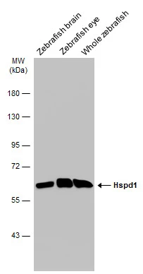
- Li L, Nan P, Zhai S, et al. Molecular cloning, characterization, and expression of hsp60 in caudal fin regeneration of Misgurnus anguillicaudatus. Mol Cell Biochem. 2014,387(1-2):143-50. doi: 10.1007/s11010-013-1879-0Read this paper
Related products
Product group Antibodies
Anti-HSP60 Antibody130-10057
ApplicationsELISA
ReactivityHuman
TargetHSPD1
- SizePrice
Product group Antibodies
References
HSP60 antibodyGTX110089
ApplicationsImmunoFluorescence, Western Blot, ELISA, ImmunoCytoChemistry, ImmunoHistoChemistry, ImmunoHistoChemistry Paraffin
ReactivityDrosophila, Hamster, Human, Mouse, Rat, Zebra Fish
TargetHSPD1
- SizePrice
Product group Antibodies
ApplicationsFlow Cytometry
TargetHSPD1
- SizePrice
Product group Antibodies
References
HSP60 Polyclonal AntibodyBS-0191R
ApplicationsImmunoFluorescence, Western Blot, ELISA, ImmunoCytoChemistry, ImmunoHistoChemistry, ImmunoHistoChemistry Frozen, ImmunoHistoChemistry Paraffin
ReactivityBovine, Canine, Equine, Human, Mouse, Rabbit, Rat
TargetHSPD1
- SizePrice
Product group Antibodies
Anti-Hsp60 AntibodyA83554
ApplicationsImmunoFluorescence, Western Blot, ELISA, ImmunoHistoChemistry
ReactivityHuman, Porcine, Rat
- SizePrice
Product group Antibodies

ApplicationsELISA
ReactivityHuman
TargetHSPD1
- SizePrice
Product group Antibodies
HSPD1 AntibodyCSB-PA002990
ApplicationsImmunoFluorescence, Western Blot, ELISA, ImmunoHistoChemistry
ReactivityHuman, Mouse, Rat
TargetHSPD1
- SizePrice

Product group Antibodies
Anti-HSPD1 AntibodyHPA001523
ApplicationsWestern Blot, ImmunoCytoChemistry, ImmunoHistoChemistry
ReactivityHuman, Mouse, Rat
TargetHSPD1
- SizePrice
Product group Antibodies

ApplicationsImmunoFluorescence, Western Blot, ELISA, ImmunoHistoChemistry
ReactivityBovine, Canine, Human, Porcine, Rat
TargetHSPD1
- SizePrice