


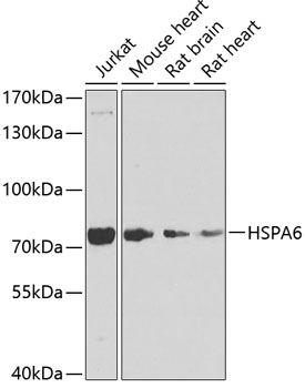
Figure 1. Western blot analysis of HSPA6 using anti-HSPA6 antibody (A05402-3). Electrophoresis was performed on a 5-20% SDS-PAGE gel at 70V (Stacking gel) / 90V (Resolving gel) for 2-3 hours. The sample well of each lane was loaded with 30 ug of sample under reducing conditions. Lane 1: human 293T whole cell lysates, Lane 2: human HL-60 whole cell lysates, Lane 3: rat testis tissue lysates, Lane 4: rat smooth muscle tissue lysates, Lane 5: rat PC-12 whole cell lysates, Lane 6: mouse testis tissue lysates, Lane 7: mouse smooth muscle tissue lysates, Lane 8: mouse Neuro-2a whole cell lysates. After electrophoresis, proteins were transferred to a nitrocellulose membrane at 150 mA for 50-90 minutes. Blocked the membrane with 5% non-fat milk/TBS for 1.5 hour at RT. The membrane was incubated with rabbit anti-HSPA6 antigen affinity purified polyclonal antibody (Catalog # A05402-3) at 0.25 microg/mL overnight at 4°C, then washed with TBS-0.1%Tween 3 times with 5 minutes each and probed with a goat anti-rabbit IgG-HRP secondary antibody at a dilution of 1:5000 for 1.5 hour at RT. The signal is developed using an Enhanced Chemiluminescent detection (ECL) kit (Catalog # EK1002) with Tanon 5200 system. A specific band was detected for HSPA6 at approximately 71 kDa. The expected band size for HSPA6 is at 71 kDa.
. HSPA6 was detected in a paraffin-embedded section of human esophageal cancer tissue. Heat mediated antigen retrieval was performed in EDTA buffer (pH 8.0, epitope retrieval solution). The tissue section was blocked with 10% goat serum. The tissue section was then incubated with 2 microg/ml rabbit anti-HSPA6 Antibody (A05402-3) overnight at 4°C. Biotinylated goat anti-rabbit IgG was used as secondary antibody and incubated for 30 minutes at 37°C. The tissue section was developed using Strepavidin-Biotin-Complex (SABC) (Catalog # SA1022) with DAB as the chromogen.")
. HSPA6 was detected in a paraffin-embedded section of human signet ring cell carcinoma of the stomach tissue. Heat mediated antigen retrieval was performed in EDTA buffer (pH 8.0, epitope retrieval solution). The tissue section was blocked with 10% goat serum. The tissue section was then incubated with 2 microg/ml rabbit anti-HSPA6 Antibody (A05402-3) overnight at 4°C. Biotinylated goat anti-rabbit IgG was used as secondary antibody and incubated for 30 minutes at 37°C. The tissue section was developed using Strepavidin-Biotin-Complex (SABC) (Catalog # SA1022) with DAB as the chromogen.")
. HSPA6 was detected in a paraffin-embedded section of human metaplasia of squamous cells of the renal pelvis tissue. Heat mediated antigen retrieval was performed in EDTA buffer (pH 8.0, epitope retrieval solution). The tissue section was blocked with 10% goat serum. The tissue section was then incubated with 2 microg/ml rabbit anti-HSPA6 Antibody (A05402-3) overnight at 4°C. Biotinylated goat anti-rabbit IgG was used as secondary antibody and incubated for 30 minutes at 37°C. The tissue section was developed using Strepavidin-Biotin-Complex (SABC) (Catalog # SA1022) with DAB as the chromogen.")
. HSPA6 was detected in a paraffin-embedded section of human tonsil tissue. Heat mediated antigen retrieval was performed in EDTA buffer (pH 8.0, epitope retrieval solution). The tissue section was blocked with 10% goat serum. The tissue section was then incubated with 2 microg/ml rabbit anti-HSPA6 Antibody (A05402-3) overnight at 4°C. Biotinylated goat anti-rabbit IgG was used as secondary antibody and incubated for 30 minutes at 37°C. The tissue section was developed using Strepavidin-Biotin-Complex (SABC) (Catalog # SA1022) with DAB as the chromogen.")
. HSPA6 was detected in an immunocytochemical section of PC-3 cells. Enzyme antigen retrieval was performed using IHC enzyme antigen retrieval reagent (AR0022) for 15 mins. The cells were blocked with 10% goat serum. And then incubated with 5 microg/mL rabbit anti-HSPA6 Antibody (A05402-3) overnight at 4°C. DyLight®488 Conjugated Goat Anti-Rabbit IgG (BA1127) was used as secondary antibody at 1:100 dilution and incubated for 30 minutes at 37°C. The section was counterstained with DAPI. Visualize using a fluorescence microscope and filter sets appropriate for the label used.")
. Overlay histogram showing Caco-2 cells stained with A05402-3 (Blue line). To facilitate intracellular staining, cells were fixed with 4% paraformaldehyde and permeabilized with permeabilization buffer. The cells were blocked with 10% normal goat serum. And then incubated with rabbit anti-HSPA6 Antibody (A05402-3, 1 microg/1x106 cells) for 30 min at 20°C. DyLight®488 conjugated goat anti-rabbit IgG (BA1127, 5-10 microg/1x106 cells) was used as secondary antibody for 30 minutes at 20°C. Isotype control antibody (Green line) was rabbit IgG (1 microg/1x106) used under the same conditions. Unlabelled sample without incubation with primary antibody and secondary antibody (Red line) was used as a blank control.")
Figure 1. Western blot analysis of HSPA6 using anti-HSPA6 antibody (A05402-3). Electrophoresis was performed on a 5-20% SDS-PAGE gel at 70V (Stacking gel) / 90V (Resolving gel) for 2-3 hours. The sample well of each lane was loaded with 30 ug of sample under reducing conditions. Lane 1: human 293T whole cell lysates, Lane 2: human HL-60 whole cell lysates, Lane 3: rat testis tissue lysates, Lane 4: rat smooth muscle tissue lysates, Lane 5: rat PC-12 whole cell lysates, Lane 6: mouse testis tissue lysates, Lane 7: mouse smooth muscle tissue lysates, Lane 8: mouse Neuro-2a whole cell lysates. After electrophoresis, proteins were transferred to a nitrocellulose membrane at 150 mA for 50-90 minutes. Blocked the membrane with 5% non-fat milk/TBS for 1.5 hour at RT. The membrane was incubated with rabbit anti-HSPA6 antigen affinity purified polyclonal antibody (Catalog # A05402-3) at 0.25 microg/mL overnight at 4°C, then washed with TBS-0.1%Tween 3 times with 5 minutes each and probed with a goat anti-rabbit IgG-HRP secondary antibody at a dilution of 1:5000 for 1.5 hour at RT. The signal is developed using an Enhanced Chemiluminescent detection (ECL) kit (Catalog # EK1002) with Tanon 5200 system. A specific band was detected for HSPA6 at approximately 71 kDa. The expected band size for HSPA6 is at 71 kDa.
Anti-HSPA6 Antibody Picoband(r)
A05402-3-CARRIER-FREE
ApplicationsFlow Cytometry, ImmunoFluorescence, Western Blot, ELISA, ImmunoCytoChemistry, ImmunoHistoChemistry
Product group Antibodies
ReactivityHuman, Mouse, Rat
TargetHSPA6
Overview
- SupplierBoster Bio
- Product NameAnti-HSPA6 Antibody Picoband(r)
- Delivery Days Customer9
- ApplicationsFlow Cytometry, ImmunoFluorescence, Western Blot, ELISA, ImmunoCytoChemistry, ImmunoHistoChemistry
- CertificationResearch Use Only
- ClonalityPolyclonal
- Concentration500 ug/ml
- Gene ID3310
- Target nameHSPA6
- Target descriptionheat shock protein family A (Hsp70) member 6
- Target synonymsHSP70B', heat shock 70 kDa protein 6, epididymis secretory sperm binding protein, heat shock 70 kDa protein B', heat shock 70kD protein 6 (HSP70B'), heat shock 70kDa protein 6 (HSP70B'), heat shock protein family A member 6
- HostRabbit
- IsotypeIgG
- Protein IDP17066
- Protein NameHeat shock 70 kDa protein 6
- Scientific DescriptionBoster Bio Anti-HSPA6 Antibody Picoband® catalog # A05402-3. Tested in ELISA, Flow Cytometry, IF, IHC, ICC, WB applications. This antibody reacts with Human, Mouse, Rat. The brand Picoband indicates this is a premium antibody that guarantees superior quality, high affinity, and strong signals with minimal background in Western blot applications. Only our best-performing antibodies are designated as Picoband, ensuring unmatched performance.
- ReactivityHuman, Mouse, Rat
- Storage Instruction-20°C,2°C to 8°C
- UNSPSC12352203
Related products
Product group Antibodies
Anti-HSPA6 AntibodyA15840
ApplicationsWestern Blot, ImmunoHistoChemistry
ReactivityHuman, Mouse, Rat
- SizePrice

Product group Antibodies
Anti-HSPA6 AntibodyHPA028549
ApplicationsImmunoHistoChemistry
ReactivityHuman
TargetHSPA6
- SizePrice
Product group Antibodies

HSPA6 / HSP70B AntibodyLS-C409241
ApplicationsWestern Blot, ImmunoHistoChemistry
ReactivityHuman, Mouse, Rat
TargetHSPA6
- SizePrice
Product group Antibodies
HSPA6 AntibodyCSB-PA863082HA01HU
ApplicationsWestern Blot, ELISA
ReactivityHuman
TargetHSPA6
- SizePrice
Product group Antibodies
HSPA6 Polyclonal AntibodyCAC13470
ApplicationsWestern Blot, ELISA
TargetHSPA6
- SizePrice
Product group Antibodies
HSPA6 antibody [C1C3]GTX106192
ApplicationsImmunoFluorescence, Western Blot, ImmunoCytoChemistry, ImmunoHistoChemistry, ImmunoHistoChemistry Paraffin
ReactivityHuman
TargetHSPA6
- SizePrice
Product group Antibodies
Anti-HSPA6 Antibody144-07688
ApplicationsWestern Blot, ImmunoHistoChemistry
ReactivityHuman, Mouse, Rat
TargetHSPA6
- SizePrice
Product group Antibodies
HSPA6 Polyclonal AntibodyBS-6540R
ApplicationsImmunoFluorescence, Western Blot, ELISA, ImmunoCytoChemistry, ImmunoHistoChemistry, ImmunoHistoChemistry Frozen, ImmunoHistoChemistry Paraffin
ReactivityBovine, Human, Mouse, Rat, Sheep
TargetHSPA6
- SizePrice