

Anti-Human CD199 Antibody, RayBright(r) Red 647
136-24082
ApplicationsFlow Cytometry
Product group Antibodies
ReactivityHuman
TargetCCR9
Overview
- SupplierRayBiotech
- Product NameAnti-Human CD199 Antibody, RayBright(r) Red 647
- Delivery Days Customer16
- ApplicationsFlow Cytometry
- CertificationResearch Use Only
- ClonalityMonoclonal
- Clone IDLS129-3C3-E3-1 [3C3]
- Concentration5 ul/test
- ConjugateOther Conjugate
- Gene ID10803
- Target nameCCR9
- Target descriptionC-C motif chemokine receptor 9
- Target synonymsCC-CKR-9, CDw199, GPR-9-6, GPR28, C-C chemokine receptor type 9, G protein-coupled receptor 28, chemokine (C-C motif) receptor 9
- HostMouse
- IsotypeIgG2b
- Protein IDP51686
- Protein NameC-C chemokine receptor type 9
- Scientific DescriptionHuman CD199 Antibody [clone: LS129-3C3-E3-1 [3C3]], monoclonal mouse IgG2b. Validated for flow cytometry.
- ReactivityHuman
- Storage Instruction2°C to 8°C
- UNSPSC12352203
Related products
Product group Antibodies
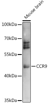
Anti-CCR9 AntibodyA91217
ApplicationsWestern Blot
ReactivityHuman, Mouse, Rat
- SizePrice
Product group Antibodies
CCR9 Recombinant AntibodyBSM-61106R
ApplicationsFlow Cytometry, ImmunoFluorescence, Western Blot, ImmunoCytoChemistry
ReactivityHuman, Mouse, Rat
TargetCCR9
- SizePrice
Product group Antibodies
Ccr9 Recombinant AntibodyCAC10227
ApplicationsImmunoFluorescence, ImmunoPrecipitation, Western Blot, ELISA
ReactivityMouse
TargetCCR9
- SizePrice
Product group Antibodies
CCR9 AntibodyCSB-PA035070
ApplicationsELISA, ImmunoHistoChemistry
ReactivityHuman, Mouse
TargetCCR9
- SizePrice

Product group Antibodies
References
ApplicationsFlow Cytometry, ImmunoFluorescence, Western Blot, ImmunoCytoChemistry
ReactivityHuman, Mouse, Rat
TargetCCR9
- SizePrice
Product group Antibodies

CCR9 / CD199 AntibodyLS-C401486
ApplicationsELISA, ImmunoHistoChemistry
ReactivityHuman, Mouse
TargetCCR9
- SizePrice

Product group Antibodies
Anti-CCR9 AntibodyHPA066879
ApplicationsImmunoCytoChemistry
ReactivityHuman
TargetCCR9
- SizePrice
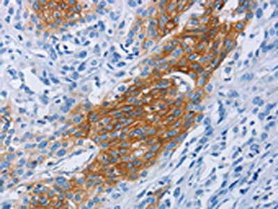
![CCR9 antibody [HL3054] detects CCR9 protein by immunohistochemical analysis. Sample: Paraffin-embedded human pancreatic cancer. CCR9 stained by CCR9 antibody [HL3054] (GTX640497) diluted at 1:100. Antigen Retrieval: Citrate buffer, pH 6.0, 15 min](https://www.genetex.com/upload/website/prouct_img/normal/GTX640497/GTX640497_T-45439_20240705_IHC-P_24070822_793.webp)
Product group Antibodies
CCR9 antibody [HL3054]GTX640497
ApplicationsFlow Cytometry, ImmunoFluorescence, Western Blot, ImmunoCytoChemistry, ImmunoHistoChemistry, ImmunoHistoChemistry Paraffin
ReactivityHuman
TargetCCR9
- SizePrice
Product group Antibodies
Anti-CCR9 AntibodyCAB14848
ApplicationsWestern Blot, ELISA
ReactivityHuman
TargetCCR9
- SizePrice