


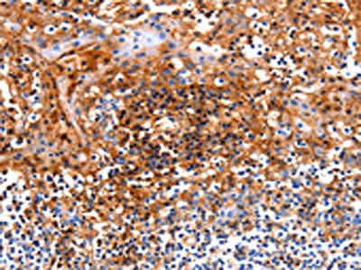
Figure 1. IHC analysis of ICOS using anti-ICOS antibody (A00291-3). ICOS was detected in paraffin-embedded section of human tonsil tissue. Heat mediated antigen retrieval was performed in citrate buffer (pH6, epitope retrieval solution) for 20 mins. The tissue section was blocked with 10% goat serum. The tissue section was then incubated with 1microg/ml rabbit anti-ICOS Antibody (A00291-3) overnight at 4°C. Biotinylated goat anti-rabbit IgG was used as secondary antibody and incubated for 30 minutes at 37°C. The tissue section was developed using Strepavidin-Biotin-Complex (SABC)(Catalog # SA1022) with DAB as the chromogen.
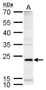
. Overlay histogram showing Raji cells stained with A00291-3 (Blue line).The cells were blocked with 10% normal goat serum. And then incubated with rabbit anti-ICOS Antibody (A00291-3,1microg/1x106 cells) for 30 min at 20°C. DyLight®488 conjugated goat anti-rabbit IgG (BA1127, 5-10microg/1x106 cells) was used as secondary antibody for 30 minutes at 20°C. Isotype control antibody (Green line) was rabbit IgG (1microg/1x106) used under the same conditions. Unlabelled sample (Red line) was also used as a control.")
Figure 1. IHC analysis of ICOS using anti-ICOS antibody (A00291-3). ICOS was detected in paraffin-embedded section of human tonsil tissue. Heat mediated antigen retrieval was performed in citrate buffer (pH6, epitope retrieval solution) for 20 mins. The tissue section was blocked with 10% goat serum. The tissue section was then incubated with 1microg/ml rabbit anti-ICOS Antibody (A00291-3) overnight at 4°C. Biotinylated goat anti-rabbit IgG was used as secondary antibody and incubated for 30 minutes at 37°C. The tissue section was developed using Strepavidin-Biotin-Complex (SABC)(Catalog # SA1022) with DAB as the chromogen.
Anti-ICOS Antibody
A00291-3-CARRIER-FREE
ApplicationsFlow Cytometry, ELISA, ImmunoCytoChemistry, ImmunoHistoChemistry
Product group Antibodies
ReactivityHuman
TargetICOS
Overview
- SupplierBoster Bio
- Product NameAnti-ICOS Antibody
- Delivery Days Customer9
- ApplicationsFlow Cytometry, ELISA, ImmunoCytoChemistry, ImmunoHistoChemistry
- CertificationResearch Use Only
- ClonalityPolyclonal
- Concentration500 ug/ml
- Gene ID29851
- Target nameICOS
- Target descriptioninducible T cell costimulator
- Target synonymsAILIM, CD278, CVID1, inducible T-cell costimulator, activation-inducible lymphocyte immunomediatory molecule, inducible T-cell co-stimulator, inducible costimulator
- HostRabbit
- IsotypeIgG
- Protein IDQ9Y6W8
- Protein NameInducible T-cell costimulator
- Scientific DescriptionBoster Bio Anti-ICOS Antibody Picoband® catalog # A00291-3. Tested in ELISA, Flow Cytometry, IHC, ICC applications. This antibody reacts with Human.
- ReactivityHuman
- Storage Instruction-20°C,2°C to 8°C
- UNSPSC12352203
Related products
Product group Antibodies
Anti-ICOS AntibodyA96181
ApplicationsWestern Blot, ELISA, ImmunoHistoChemistry
ReactivityHuman, Mouse, Rat
- SizePrice
Product group Antibodies
Anti-ICOS Antibody144-01811
ApplicationsImmunoFluorescence, Western Blot, ImmunoHistoChemistry
ReactivityHuman, Mouse, Rat
TargetICOS
- SizePrice
Product group Antibodies
Anti-ICOS AntibodyAMAB91995
ApplicationsWestern Blot
ReactivityHuman
TargetICOS
- SizePrice
Product group Antibodies
ICOS Recombinant AntibodyBSM-60227R
ApplicationsFlow Cytometry, ImmunoFluorescence, Western Blot, ImmunoCytoChemistry, ImmunoHistoChemistry, ImmunoHistoChemistry Paraffin
ReactivityHuman
TargetICOS
- SizePrice
Product group Antibodies
ICOS AntibodyCSB-PA123586
ApplicationsELISA, ImmunoHistoChemistry
ReactivityHuman
TargetICOS
- SizePrice
Product group Antibodies

Goat anti-ICOSEB09484
ApplicationsELISA, ImmunoHistoChemistry
ReactivityHuman
TargetICOS
- SizePrice
Product group Antibodies
ApplicationsImmunoPrecipitation, Western Blot, ImmunoCytoChemistry, ImmunoHistoChemistry
TargetICOS
- SizePrice
Product group Antibodies

ICOS / CD278 Antibody (Internal)LS-C368597
ApplicationsWestern Blot
ReactivityHuman, Mouse, Rat
TargetICOS
- SizePrice
Product group Antibodies
ICOS antibodyGTX129570
ApplicationsWestern Blot
ReactivityHuman
TargetICOS
- SizePrice