Bio-Connect
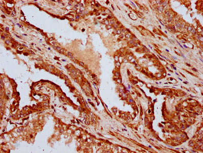
Immunohistochemical staining of human caudate shows strong cytoplasmic positivity in astrocytes.

Anti-IFT172 Antibody

HPA044893
Atlas Antibodies
ApplicationsImmunoHistoChemistry
Product group Antibodies
TargetIFT172
Sign in to order and to see your custom pricing.
Large volume orders?
Order with a bulk request

Overview

  • Supplier
    Atlas Antibodies
  • Product Name
    Anti-IFT172
  • Delivery Days Customer
    4
  • Applications
    ImmunoHistoChemistry
  • Certification
    Research Use Only
  • Clonality
    Polyclonal
  • Conjugate
    Unconjugated
  • Gene ID26160
  • Target name
    IFT172
  • Target description
    intraflagellar transport 172
  • Target synonyms
    BBS20; intraflagellar transport 172 homolog; intraflagellar transport protein 172 homolog; NPHP17; osm-1; RP71; selective LIM binding factor homolog; SLB; SRTD10; wim; wimple homolog
  • Host
    Rabbit
  • Isotype
    IgG
  • Protein IDQ9UG01
  • Protein Name
    Intraflagellar transport protein 172 homolog
  • Scientific Description
    Recombinant Protein Epitope Signature Tag (PrEST) antigen sequence
  • Storage Instruction
    -20°C,2°C to 8°C
  • UNSPSC
    12352203

Related products

IHC image of CSB-PA887019LA01HU diluted at 1:400 and staining in paraffin-embedded human prostate cancer performed on a Leica BondTM system. After dewaxing and hydration, antigen retrieval was mediated by high pressure in a citrate buffer (pH 6.0). Section was blocked with 10% normal goat serum 30min at RT. Then primary antibody (1% BSA) was incubated at 4°C overnight. The primary is detected by a biotinylated secondary antibody and visualized using an HRP conjugated SP system.
Product group Antibodies
Cusabio
IFT172 AntibodyCSB-PA887019LA01HU
ApplicationsImmunoFluorescence, ELISA, ImmunoHistoChemistry
TargetIFT172
  • SizePrice
Figure 1. Western blot analysis of IFT172 using anti-IFT172 antibody (A08217-1). Electrophoresis was performed on a 5-20% SDS-PAGE gel at 70V (Stacking gel) / 90V (Resolving gel) for 2-3 hours. The sample well of each lane was loaded with 30 ug of sample under reducing conditions. Lane 1: human A549 whole cell lysates, Lane 2: human HEL whole cell lysates, Lane 3: human U251 whole cell lysates, Lane 4: human 293T whole cell lysates, Lane 5: rat testis tissue lysates, Lane 6: rat brain tissue lysates, Lane 7: mouse testis tissue lysates, Lane 8: mouse brain tissue lysates. After electrophoresis, proteins were transferred to a nitrocellulose membrane at 150 mA for 50-90 minutes. Blocked the membrane with 5% non-fat milk/TBS for 1.5 hour at RT. The membrane was incubated with rabbit anti-IFT172 antigen affinity purified polyclonal antibody (Catalog # A08217-1) at 0.5 microg/mL overnight at 4°C, then washed with TBS-0.1%Tween 3 times with 5 minutes each and probed with a goat anti-rabbit IgG-HRP secondary antibody at a dilution of 1:5000 for 1.5 hour at RT. The signal is developed using an Enhanced Chemiluminescent detection (ECL) kit (Catalog # EK1002) with Tanon 5200 system. A specific band was detected for IFT172 at approximately 180 kDa. The expected band size for IFT172 is at 198 kDa.
Product group Antibodies
Boster Bio
ApplicationsWestern Blot, ELISA
TargetIFT172
  • SizePrice
Atlas Antibodies
Protein IDQ9UG01
  • SizePrice
Product group Antibodies
LifeSpan BioSciences
ApplicationsELISA
TargetIFT172
  • SizePrice
Product group Antibodies
Assay Genie
ApplicationsImmunoFluorescence, ELISA, ImmunoHistoChemistry
TargetIFT172
  • SizePrice