


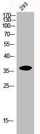
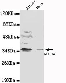
Figure 1. Western blot analysis of IKB Alpha using anti-IKB Alpha antibody (PB9291). Electrophoresis was performed on a 5-20% SDS-PAGE gel at 70V (Stacking gel) / 90V (Resolving gel) for 2-3 hours. The sample well of each lane was loaded with 50ug of sample under reducing conditions. Lane 1: human Jurkat whole cell lysates, Lane 2: human MDA-MB-453 whole cell lysates, Lane 3: human SW620 whole cell lysates, Lane 4: human A549 whole cell lysates. After Electrophoresis, proteins were transferred to a Nitrocellulose membrane at 150mA for 50-90 minutes. Blocked the membrane with 5% Non-fat Milk/ TBS for 1.5 hour at RT. The membrane was incubated with rabbit anti-IKB Alpha antigen affinity purified polyclonal antibody (Catalog # PB9291) at 0.5 microg/mL overnight at 4°C, then washed with TBS-0.1%Tween 3 times with 5 minutes each and probed with a goat anti-rabbit IgG-HRP secondary antibody at a dilution of 1:10000 for 1.5 hour at RT. The signal is developed using an Enhanced Chemiluminescent detection (ECL) kit (Catalog # EK1002) with Tanon 5200 system. A specific band was detected for IKB Alpha at approximately 39KD. The expected band size for IKB Alpha is at 36KD.
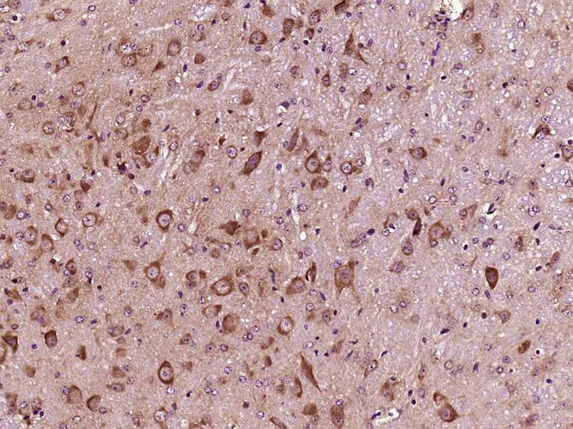
. IKB alpha was detected in paraffin-embedded section of Mouse Intestine Tissue. Heat mediated antigen retrieval was performed in citrate buffer (pH6, epitope retrieval solution) for 20 mins. The tissue section was blocked with 10% goat serum. The tissue section was then incubated with 1microg/ml rabbit anti-IKB alpha Antibody (PB9291) overnight at 4°C. Biotinylated goat anti-rabbit IgG was used as secondary antibody and incubated for 30 minutes at 37°C. The tissue section was developed using Strepavidin-Biotin-Complex (SABC)(Catalog # SA1022) with DAB as the chromogen.")
. IKB alpha was detected in paraffin-embedded section of Rat Intestine Tissue. Heat mediated antigen retrieval was performed in citrate buffer (pH6, epitope retrieval solution) for 20 mins. The tissue section was blocked with 10% goat serum. The tissue section was then incubated with 1microg/ml rabbit anti-IKB alpha Antibody (PB9291) overnight at 4°C. Biotinylated goat anti-rabbit IgG was used as secondary antibody and incubated for 30 minutes at 37°C. The tissue section was developed using Strepavidin-Biotin-Complex (SABC)(Catalog # SA1022) with DAB as the chromogen.")
. IKB alpha was detected in paraffin-embedded section of Human Mammary Cancer Tissue. Heat mediated antigen retrieval was performed in citrate buffer (pH6, epitope retrieval solution) for 20 mins. The tissue section was blocked with 10% goat serum. The tissue section was then incubated with 1microg/ml rabbit anti-IKB alpha Antibody (PB9291) overnight at 4°C. Biotinylated goat anti-rabbit IgG was used as secondary antibody and incubated for 30 minutes at 37°C. The tissue section was developed using Strepavidin-Biotin-Complex (SABC)(Catalog # SA1022) with DAB as the chromogen.")
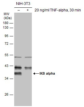
. Electrophoresis was performed on a 5-20% SDS-PAGE gel at 70V (Stacking gel) / 90V (Resolving gel) for 2-3 hours. The sample well of each lane was loaded with 50ug of sample under reducing conditions. Lane 1: rat brain tissue lysates Lane 2: rat lung tissue lysates Lane 3: rat heart tissue lysates Lane 4: rat skeletal muscle tissue lysates Lane 5: mouse lung tissue lysates Lane 6: mouse heart tissue lysates After Electrophoresis, proteins were transferred to a Nitrocellulose membrane at 150mA for 50-90 minutes. Blocked the membrane with 5% Non-fat Milk/ TBS for 1.5 hour at RT. The membrane was incubated with rabbit anti-IKB Alpha antigen affinity purified polyclonal antibody (Catalog # PB9291) at 0.5 microg/mL overnight at 4°C, then washed with TBS-0.1%Tween 3 times with 5 minutes each and probed with a goat anti-rabbit IgG-HRP secondary antibody at a dilution of 1:10000 for 1.5 hour at RT. The signal is developed using an Enhanced Chemiluminescent detection (ECL) kit (Catalog # EK1002) with Tanon 5200 system. A specific band was detected for IKB Alpha at approximately 39KD. The expected band size for IKB Alpha is at 36KD.")
Figure 1. Western blot analysis of IKB Alpha using anti-IKB Alpha antibody (PB9291). Electrophoresis was performed on a 5-20% SDS-PAGE gel at 70V (Stacking gel) / 90V (Resolving gel) for 2-3 hours. The sample well of each lane was loaded with 50ug of sample under reducing conditions. Lane 1: human Jurkat whole cell lysates, Lane 2: human MDA-MB-453 whole cell lysates, Lane 3: human SW620 whole cell lysates, Lane 4: human A549 whole cell lysates. After Electrophoresis, proteins were transferred to a Nitrocellulose membrane at 150mA for 50-90 minutes. Blocked the membrane with 5% Non-fat Milk/ TBS for 1.5 hour at RT. The membrane was incubated with rabbit anti-IKB Alpha antigen affinity purified polyclonal antibody (Catalog # PB9291) at 0.5 microg/mL overnight at 4°C, then washed with TBS-0.1%Tween 3 times with 5 minutes each and probed with a goat anti-rabbit IgG-HRP secondary antibody at a dilution of 1:10000 for 1.5 hour at RT. The signal is developed using an Enhanced Chemiluminescent detection (ECL) kit (Catalog # EK1002) with Tanon 5200 system. A specific band was detected for IKB Alpha at approximately 39KD. The expected band size for IKB Alpha is at 36KD.
Anti-IKB alpha/NFKBIA Antibody Picoband(r)
PB9291-CARRIER-FREE
ApplicationsWestern Blot
Product group Antibodies
ReactivityHuman
TargetNFKBIA
Overview
- SupplierBoster Bio
- Product NameAnti-IKB alpha/NFKBIA Antibody Picoband(r)
- Delivery Days Customer9
- Application Supplier NoteWB: The detection limit for IKB alpha is approximately 0.2ng/lane under reducing conditions. Tested Species: In-house tested species with positive results. By Heat: Boiling the paraffin sections in 10mM citrate buffer, pH6.0, for 20mins is required for the staining of formalin/paraffin sections. Other applications have not been tested. Optimal dilutions should be determined by end users.
- ApplicationsWestern Blot
- CertificationResearch Use Only
- ClonalityPolyclonal
- Concentration500 ug/ml
- Gene ID4792
- Target nameNFKBIA
- Target descriptionNFKB inhibitor alpha
- Target synonymsEDAID2, IKBA, MAD-3, NFKBI, NF-kappa-B inhibitor alpha, I-kappa-B-alpha, IkappaBalpha, ikB-alpha, major histocompatibility complex enhancer-binding protein MAD3, nuclear factor of kappa light chain gene enhancer in B-cells, nuclear factor of kappa light polypeptide gene enhancer in B-cells inhibitor, alpha
- HostRabbit
- IsotypeIgG
- Protein IDP25963
- Protein NameNF-kappa-B inhibitor alpha
- Scientific DescriptionBoster Bio Anti-IKB alpha/NFKBIA Antibody Picoband® catalog # PB9291. Tested in WB applications. This antibody reacts with Human. The brand Picoband indicates this is a premium antibody that guarantees superior quality, high affinity, and strong signals with minimal background in Western blot applications. Only our best-performing antibodies are designated as Picoband, ensuring unmatched performance.
- ReactivityHuman
- Storage Instruction-20°C,2°C to 8°C
- UNSPSC12352203
Related products
Product group Antibodies
NFKBIA AntibodyCSB-PA003073
ApplicationsWestern Blot, ELISA
ReactivityHuman, Mouse, Rat
TargetNFKBIA
- SizePrice
Product group Antibodies
ApplicationsWestern Blot
ReactivityHuman
- SizePrice
Product group Antibodies

NFKBIA / IKB Alpha / IKBA AntibodyLS-C814109
ApplicationsWestern Blot
ReactivityHuman, Mouse, Porcine, Rat, Sheep
TargetNFKBIA
- SizePrice

Product group Antibodies
Anti-NFKBIA AntibodyHPA029207
ApplicationsWestern Blot, ImmunoCytoChemistry, ImmunoHistoChemistry
ReactivityHuman
TargetNFKBIA
- SizePrice
Product group Antibodies
NFKBIA Polyclonal AntibodyCAC15883
ApplicationsImmunoFluorescence, Western Blot, ELISA, ImmunoHistoChemistry
TargetNFKBIA
- SizePrice
Product group Antibodies
References
ApplicationsImmunoFluorescence, Western Blot, ELISA, ImmunoCytoChemistry, ImmunoHistoChemistry, ImmunoHistoChemistry Frozen, ImmunoHistoChemistry Paraffin
ReactivityBovine, Human, Mouse, Porcine, Rabbit, Rat, Sheep
TargetNFKBIA
- SizePrice
Product group Antibodies
IKB alpha antibodyGTX110521
ApplicationsImmunoFluorescence, ImmunoPrecipitation, Western Blot, ImmunoCytoChemistry, ImmunoHistoChemistry, ImmunoHistoChemistry Paraffin
ReactivityHuman, Mouse, Porcine
TargetNFKBIA
- SizePrice
Product group Antibodies
TargetNFKBIA
- SizePrice