

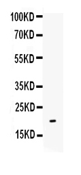
Figure. Western blot analysis of IL-10 using anti-IL-10 antibody (RP1014). Electrophoresis was performed on a 5-20% SDS-PAGE gel at 70V (Stacking gel) / 90V (Resolving gel) for 2-3 hours. The sample well of each lane was loaded with 50ug of sample under reducing conditions. Lane : Recombinant Human IL-10 Protein 0.5ng After Electrophoresis, proteins were transferred to a Nitrocellulose membrane at 150mA for 50-90 minutes. Blocked the membrane with 5% Non-fat Milk/ TBS for 1.5 hour at RT. The membrane was incubated with rabbit anti-IL-10 antigen affinity purified polyclonal antibody (Catalog # RP1014) at 0.5 microg/mL overnight at 4°C, then washed with TBS-0.1%Tween 3 times with 5 minutes each and probed with a goat anti-rabbit IgG-HRP secondary antibody at a dilution of 1:10000 for 1.5 hour at RT. The signal is developed using an Enhanced Chemiluminescent detection (ECL) kit (Catalog # EK1002) with Tanon 5200 system. A specific band was detected for IL-10 at approximately 19KD. The expected band size for IL-10 is at 19KD.
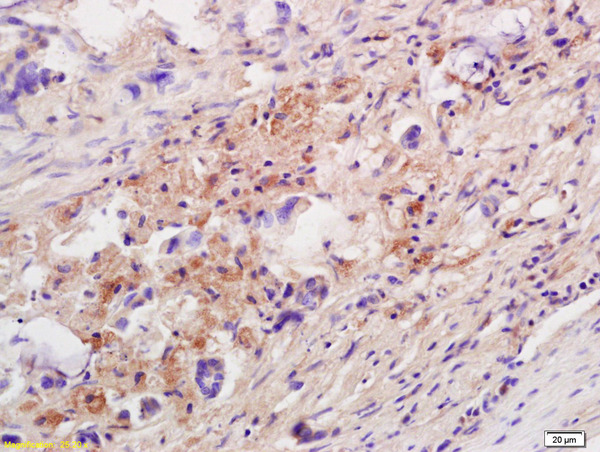
. IL10 was detected in paraffin-embedded section of human appendicitis tissue. Heat mediated antigen retrieval was performed in EDTA buffer (pH8.0, epitope retrieval solution). The tissue section was blocked with 10% goat serum. The tissue section was then incubated with 1microg/ml rabbit anti-IL10 Antibody (RP1014) overnight at 4°C. Biotinylated goat anti-rabbit IgG was used as secondary antibody and incubated for 30 minutes at 37°C. The tissue section was developed using Strepavidin-Biotin-Complex (SABC) (Catalog # SA1022) with DAB as the chromogen.")
Figure. Western blot analysis of IL-10 using anti-IL-10 antibody (RP1014). Electrophoresis was performed on a 5-20% SDS-PAGE gel at 70V (Stacking gel) / 90V (Resolving gel) for 2-3 hours. The sample well of each lane was loaded with 50ug of sample under reducing conditions. Lane : Recombinant Human IL-10 Protein 0.5ng After Electrophoresis, proteins were transferred to a Nitrocellulose membrane at 150mA for 50-90 minutes. Blocked the membrane with 5% Non-fat Milk/ TBS for 1.5 hour at RT. The membrane was incubated with rabbit anti-IL-10 antigen affinity purified polyclonal antibody (Catalog # RP1014) at 0.5 microg/mL overnight at 4°C, then washed with TBS-0.1%Tween 3 times with 5 minutes each and probed with a goat anti-rabbit IgG-HRP secondary antibody at a dilution of 1:10000 for 1.5 hour at RT. The signal is developed using an Enhanced Chemiluminescent detection (ECL) kit (Catalog # EK1002) with Tanon 5200 system. A specific band was detected for IL-10 at approximately 19KD. The expected band size for IL-10 is at 19KD.
Anti-Interleukin-10 IL10 Antibody Picoband(r)
RP1014
ApplicationsWestern Blot, ELISA, ImmunoHistoChemistry
Product group Antibodies
ReactivityHuman
TargetIL10
Overview
- SupplierBoster Bio
- Product NameAnti-Interleukin-10 IL10 Antibody Picoband(r)
- Delivery Days Customer9
- Application Supplier NoteBy Heat: Boiling the paraffin sections in 10mM citrate buffer, pH6.0, for 20mins is required for the staining of formalin/paraffin sections. Other applications have not been tested. Optimal dilutions should be determined by end users.
- ApplicationsWestern Blot, ELISA, ImmunoHistoChemistry
- Applications SupplierELI, IHP, WB, IHC
- CertificationResearch Use Only
- ClonalityPolyclonal
- Concentration500 ug/ml
- Gene ID3586
- Target nameIL10
- Target descriptioninterleukin 10
- Target synonymsCSIF, GVHDS, IL-10, IL10A, TGIF, interleukin-10, T-cell growth inhibitory factor, cytokine synthesis inhibitory factor
- HostRabbit
- IsotypeIgG
- Protein IDP22301
- Protein NameInterleukin-10
- Scientific DescriptionBoster Bio Anti-Interleukin-10 IL10 Antibody catalog # RP1014. Tested in ELISA, IHC, WB applications. This antibody reacts with Human. The brand Picoband indicates this is a premium antibody that guarantees superior quality, high affinity, and strong signals with minimal background in Western blot applications. Only our best-performing antibodies are designated as Picoband, ensuring unmatched performance.
- ReactivityHuman
- Reactivity SupplierHuman
- Storage Instruction-20°C,2°C to 8°C
- UNSPSC12352203
References
- Zhao B, Gao WW, Liu YJ, et al. The role of glycogen synthase kinase 3 beta in brain injury induced by myocardial ischemia/reperfusion injury in a rat model of diabetes mellitus. Neural Regen Res. 2017,12(10):1632-1639. doi: 10.4103/1673-5374.217337Read this paper
- Wang LQ, Yan XT, Yan CF, et al. Women with Recurrent Miscarriage Have Decreased Expression of 25-Hydroxyvitamin D3-1α-Hydroxylase by the Fetal-Maternal Interface. PLoS One. 2016,11(12):e0165589. doi: 10.1371/journal.pone.0165589Read this paper
- Zhu XW, Zhu HZ, Zhu YQ, et al. Foxp3 expression in CD4(+)CD25(+)Foxp3(+) regulatory T cells promotes development of colorectal cancer by inhibiting tumor immunity. J Huazhong Univ Sci Technolog Med Sci. 2016,36(5):677-682. doi: 10.1007/s11596-016-1644-1Read this paper
- Li G, Ren J, Wang G, et al. T2 enhances in situ level of Foxp3+ regulatory cells and modulates inflammatory cytokines in Crohn's disease. Int Immunopharmacol. 2014,18(2):244-8. doi: 10.1016/j.intimp.2013.12.014Read this paper
- Wang F, Li H, Yang Z, et al. Expression of interleukin-10 in patients with adenomyosis. Fertil Steril. 2009,91(5):1681-5. doi: 10.1016/j.fertnstert.2008.02.164Read this paper
Datasheet
MSDS
Related products
Product group Antibodies

Anti-IL-10 [JES3-9D7]Ab00139-23.0
ApplicationsWestern Blot, ELISA, ELISpot Assay, ImmunoHistoChemistry, ImmunoHistoChemistry Paraffin, Neutralisation/Blocking
ReactivityHuman, Monkey, Primate
TargetIL10
- SizePrice

Product group Antibodies
ApplicationsELISA, ELISpot Assay
ReactivityHuman
TargetIL10
- SizePrice
Product group Antibodies
Anti-IL10 AntibodyA101419
ApplicationsWestern Blot, ELISA
ReactivityHuman
- SizePrice
Product group Antibodies

IL-10 Antibody (clone JES5-16E3, FITC)LS-C810819
ApplicationsFlow Cytometry
ReactivityMouse
TargetIL10
- SizePrice
Product group Antibodies
References
IL-10 Polyclonal AntibodyBS-6761R
ApplicationsImmunoFluorescence, Western Blot, ELISA, ImmunoCytoChemistry, ImmunoHistoChemistry, ImmunoHistoChemistry Frozen, ImmunoHistoChemistry Paraffin
ReactivityChicken, Guinea Pig, Human, Mouse, Porcine, Rabbit, Rat, Sheep
TargetIL10
- SizePrice

Product group Antibodies
Anti-IL10 Antibody Picoband(r)A00021-2-CARRIER-FREE
ApplicationsWestern Blot, ELISA
ReactivityHuman, Mouse, Rat
TargetIL10
- SizePrice
Product group Antibodies
ApplicationsImmunoPrecipitation, Western Blot, ImmunoCytoChemistry, ImmunoHistoChemistry
TargetIL10
- SizePrice
Product group Antibodies
IL10 AntibodyCSB-PA004870
ApplicationsWestern Blot, ELISA
ReactivityHuman
TargetIL10
- SizePrice