


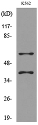
Figure 1. Western blot analysis of IL2 Receptor gamma/IL2RG using anti-IL2 Receptor gamma/IL2RG antibody (A00308-1). Electrophoresis was performed on a 5-20% SDS-PAGE gel at 70V (Stacking gel) / 90V (Resolving gel) for 2-3 hours. The sample well of each lane was loaded with 30ug of sample under reducing conditions. Lane 1: human Jurkat whole cell lysates, Lane 2: human K562 whole cell lysates, Lane 3: human Raji whole cell lysates, Lane 4: human U-937 whole cell lysates, Lane 5: human HEK293 whole cell lysates, Lane 6: human HMY2.CIR whole cell lysates, Lane 7: human THP-1 whole cell lysates, Lane 8: rat RH-35 whole cell lysates, Lane 9: rat C6 whole cell lysates, Lane 10: mouse HEPA1-6 whole cell lysates. After Electrophoresis, proteins were transferred to a Nitrocellulose membrane at 150mA for 50-90 minutes. Blocked the membrane with 5% Non-fat Milk/ TBS for 1.5 hour at RT. The membrane was incubated with rabbit anti-IL2 Receptor gamma/IL2RG antigen affinity purified polyclonal antibody (Catalog # A00308-1) at 0.5 microg/mL overnight at 4°C, then washed with TBS-0.1%Tween 3 times with 5 minutes each and probed with a goat anti-rabbit IgG-HRP secondary antibody at a dilution of 1:5000 for 1.5 hour at RT. The signal is developed using an Enhanced Chemiluminescent detection (ECL) kit (Catalog # EK1002) with Tanon 5200 system. A specific band was detected for IL2 Receptor gamma/IL2RG at approximately 65KD. The expected band size for IL2 Receptor gamma/IL2RG is at 65KD.
. IL2 Receptor gamma/IL2RG was detected in immunocytochemical section of K562 cells. Enzyme antigen retrieval was performed using IHC enzyme antigen retrieval reagent (AR0022) for 15 mins. The cells were blocked with 10% goat serum. And then incubated with 5microg/mL rabbit anti-IL2 Receptor gamma/IL2RG Antibody (A00308-1) overnight at 4°C. DyLight®594 Conjugated Goat Anti-Rabbit IgG (BA1142) was used as secondary antibody at 1:100 dilution and incubated for 30 minutes at 37°C. The section was counterstained with DAPI. Visualize using a fluorescence microscope and filter sets appropriate for the label used.")
Figure 1. Western blot analysis of IL2 Receptor gamma/IL2RG using anti-IL2 Receptor gamma/IL2RG antibody (A00308-1). Electrophoresis was performed on a 5-20% SDS-PAGE gel at 70V (Stacking gel) / 90V (Resolving gel) for 2-3 hours. The sample well of each lane was loaded with 30ug of sample under reducing conditions. Lane 1: human Jurkat whole cell lysates, Lane 2: human K562 whole cell lysates, Lane 3: human Raji whole cell lysates, Lane 4: human U-937 whole cell lysates, Lane 5: human HEK293 whole cell lysates, Lane 6: human HMY2.CIR whole cell lysates, Lane 7: human THP-1 whole cell lysates, Lane 8: rat RH-35 whole cell lysates, Lane 9: rat C6 whole cell lysates, Lane 10: mouse HEPA1-6 whole cell lysates. After Electrophoresis, proteins were transferred to a Nitrocellulose membrane at 150mA for 50-90 minutes. Blocked the membrane with 5% Non-fat Milk/ TBS for 1.5 hour at RT. The membrane was incubated with rabbit anti-IL2 Receptor gamma/IL2RG antigen affinity purified polyclonal antibody (Catalog # A00308-1) at 0.5 microg/mL overnight at 4°C, then washed with TBS-0.1%Tween 3 times with 5 minutes each and probed with a goat anti-rabbit IgG-HRP secondary antibody at a dilution of 1:5000 for 1.5 hour at RT. The signal is developed using an Enhanced Chemiluminescent detection (ECL) kit (Catalog # EK1002) with Tanon 5200 system. A specific band was detected for IL2 Receptor gamma/IL2RG at approximately 65KD. The expected band size for IL2 Receptor gamma/IL2RG is at 65KD.
Anti-IL2 Receptor gamma/IL2RG Antibody Picoband(r)
A00308-1-CARRIER-FREE
ApplicationsImmunoFluorescence, Western Blot, ImmunoCytoChemistry
Product group Antibodies
ReactivityHuman, Mouse, Rat
TargetIL2RG
Overview
- SupplierBoster Bio
- Product NameAnti-IL2 Receptor gamma/IL2RG Antibody Picoband(r)
- Delivery Days Customer9
- ApplicationsImmunoFluorescence, Western Blot, ImmunoCytoChemistry
- CertificationResearch Use Only
- ClonalityPolyclonal
- Concentration500 ug/ml
- Gene ID3561
- Target nameIL2RG
- Target descriptioninterleukin 2 receptor subunit gamma
- Target synonymsCD132, CIDX, IL-2RG, IMD4, P64, SCIDX, SCIDX1, cytokine receptor common subunit gamma, CD132 antigen, IL-2 receptor subunit gamma, IL-2R subunit gamma, common cytokine receptor gamma chain, gammaC, interleukin 2 receptor, gamma, nonfunctional common cytokine receptor gamma chain
- HostRabbit
- IsotypeIgG
- Protein IDP31785
- Protein NameCytokine receptor common subunit gamma
- Scientific DescriptionBoster Bio Anti-IL2 Receptor gamma/IL2RG Antibody Picoband® catalog # A00308-1. Tested in IF, ICC, WB applications. This antibody reacts with Human, Mouse, Rat. The brand Picoband indicates this is a premium antibody that guarantees superior quality, high affinity, and strong signals with minimal background in Western blot applications. Only our best-performing antibodies are designated as Picoband, ensuring unmatched performance.
- ReactivityHuman, Mouse, Rat
- Storage Instruction-20°C,2°C to 8°C
- UNSPSC12352203
References
- Wang X, Rickert M, Garcia KC. Structure of the quaternary complex of interleukin-2 with its alpha, beta, and gammac receptors. Science. 2005,310(5751):1159-63.Read this paper
Related products
Product group Antibodies
Anti-IL2RG AntibodyA101019
ApplicationsWestern Blot, ELISA
ReactivityHuman
- SizePrice
Product group Antibodies
Anti-IL2RG Antibody144-65354
ApplicationsWestern Blot, ImmunoHistoChemistry
ReactivityHuman, Mouse
TargetIL2RG
- SizePrice
Product group Antibodies

IL2RG / CD132 Antibody (clone 3E12)LS-C770033
ApplicationsFlow Cytometry
ReactivityMouse
TargetIL2RG
- SizePrice
Product group Antibodies
IL-2R gamma Polyclonal AntibodyBS-2545R
ApplicationsFlow Cytometry, Western Blot, ELISA
ReactivityBovine, Canine, Human, Mouse, Porcine, Rat
TargetIL2RG
- SizePrice
Product group Antibodies
IL2RG Polyclonal AntibodyCAC13780
ApplicationsWestern Blot, ELISA, ImmunoHistoChemistry
TargetIL2RG
- SizePrice
Product group Antibodies
IL2RG AntibodyCSB-PA005615
ApplicationsWestern Blot, ELISA
ReactivityHuman
TargetIL2RG
- SizePrice
Product group Antibodies
IL2 Receptor gamma antibodyGTX54363
ApplicationsWestern Blot
ReactivityHuman, Mouse
TargetIL2RG
- SizePrice
Product group Antibodies
Anti-TXLNA AntibodyCAB18291
ApplicationsImmunoFluorescence, Western Blot, ELISA, ImmunoCytoChemistry
ReactivityHuman
TargetIL2RG
- SizePrice