


Immunohistochemical staining of human Stomach shows strong cytoplasmic and membranous positivity in glandular cells.
Immunohistochemical staining of human Stomach shows strong cytoplasmic and membranous positivity in glandular cells.
Anti-IL7 Antibody
HPA019590
ApplicationsImmunoHistoChemistry
Product group Antibodies
ReactivityHuman
TargetIL7
Overview
- SupplierAtlas Antibodies
- Product NameAnti-IL7 Antibody
- Delivery Days Customer4
- ApplicationsImmunoHistoChemistry
- CertificationResearch Use Only
- ClonalityPolyclonal
- ConjugateUnconjugated
- Gene ID3574
- Target nameIL7
- Target descriptioninterleukin 7
- Target synonymsIL-7, IMD130, interleukin-7
- HostRabbit
- IsotypeIgG
- Protein IDP13232
- Protein NameInterleukin-7
- Scientific DescriptionRecombinant Protein Epitope Signature Tag (PrEST) antigen sequence
- ReactivityHuman
- Storage Instruction-20°C,2°C to 8°C
- UNSPSC41116161
Datasheet
MSDS
Related products
Product group Antibodies
IL7 AntibodyCSB-PA011669KA01HU
ApplicationsWestern Blot, ELISA, ImmunoHistoChemistry
ReactivityHuman, Mouse, Rat
TargetIL7
- SizePrice

Product group Antibodies
Anti-IL7 AntibodyAMAB91684
ApplicationsImmunoCytoChemistry
ReactivityHuman
TargetIL7
- SizePrice
Product group Antibodies
Anti-IL7 AntibodyAMAB91684
ApplicationsImmunoCytoChemistry
ReactivityHuman
TargetIL7
- SizePrice
Product group Antibodies
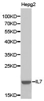
Anti-IL7 AntibodyA28549
ApplicationsWestern Blot
ReactivityHuman, Mouse, Rat
- SizePrice
Product group Antibodies

IL7 AntibodyLS-C831251
ApplicationsELISA, ImmunoHistoChemistry
ReactivityHuman
TargetIL7
- SizePrice
Product group Antibodies
ApplicationsImmunoPrecipitation, Western Blot, ImmunoCytoChemistry, ImmunoHistoChemistry
TargetIL7
- SizePrice

Product group Antibodies
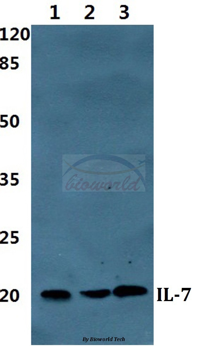
Anti-IL7 Antibody Picoband(r)PB9521-CARRIER-FREE
ApplicationsWestern Blot, ELISA
ReactivityHuman
TargetIL7
- SizePrice