


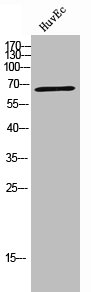
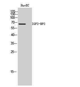
Figure 1. Western blot analysis of IMP2 using anti-IMP2 antibody (M02010-1). Electrophoresis was performed on a 5-20% SDS-PAGE gel at 70V (Stacking gel) / 90V (Resolving gel) for 2-3 hours. The sample well of each lane was loaded with 30 ug of sample under reducing conditions. Lane 1: human SH-SY5Y whole cell lysates, Lane 2: human Hela whole cell lysates, Lane 3: human HepG2 whole cell lysates, Lane 4: human A431 whole cell lysates, Lane 5: rat heart tissue lysates, Lane 6: mouse heart tissue lysates, Lane 7: mouse NIH/3T3 whole cell lysates. After electrophoresis, proteins were transferred to a nitrocellulose membrane at 150 mA for 50-90 minutes. Blocked the membrane with 5% non-fat milk/TBS for 1.5 hour at RT. The membrane was incubated with rabbit anti-IMP2 antigen affinity purified monoclonal antibody (Catalog # M02010-1) at 1:500 overnight at 4°C, then washed with TBS-0.1%Tween 3 times with 5 minutes each and probed with a goat anti-rabbit IgG-HRP secondary antibody at a dilution of 1:1000 for 1.5 hour at RT. The signal is developed using an Enhanced Chemiluminescent detection (ECL) kit (Catalog # EK1002) with Tanon 5200 system. A specific band was detected for IMP2 at approximately 66 kDa. The expected band size for IMP2 is at 66 kDa.
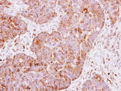
. IMP2 was detected in a paraffin-embedded section of human lung cancer tissue. Heat mediated antigen retrieval was performed in EDTA buffer (pH 8.0, epitope retrieval solution). The tissue section was blocked with 10% goat serum. The tissue section was then incubated with 1:50 rabbit anti-IMP2 Antibody (M02010-1) overnight at 4°C. Peroxidase Conjugated Goat Anti-rabbit IgG was used as secondary antibody and incubated for 30 minutes at 37°C. The tissue section was developed using HRP Conjugated Rabbit IgG Super Vision Assay Kit (Catalog # SV0002) with DAB as the chromogen.")
. IMP2 was detected in a paraffin-embedded section of human testicular germ cell tumor tissue. Heat mediated antigen retrieval was performed in EDTA buffer (pH 8.0, epitope retrieval solution). The tissue section was blocked with 10% goat serum. The tissue section was then incubated with 1:50 rabbit anti-IMP2 Antibody (M02010-1) overnight at 4°C. Peroxidase Conjugated Goat Anti-rabbit IgG was used as secondary antibody and incubated for 30 minutes at 37°C. The tissue section was developed using HRP Conjugated Rabbit IgG Super Vision Assay Kit (Catalog # SV0002) with DAB as the chromogen.")
Figure 1. Western blot analysis of IMP2 using anti-IMP2 antibody (M02010-1). Electrophoresis was performed on a 5-20% SDS-PAGE gel at 70V (Stacking gel) / 90V (Resolving gel) for 2-3 hours. The sample well of each lane was loaded with 30 ug of sample under reducing conditions. Lane 1: human SH-SY5Y whole cell lysates, Lane 2: human Hela whole cell lysates, Lane 3: human HepG2 whole cell lysates, Lane 4: human A431 whole cell lysates, Lane 5: rat heart tissue lysates, Lane 6: mouse heart tissue lysates, Lane 7: mouse NIH/3T3 whole cell lysates. After electrophoresis, proteins were transferred to a nitrocellulose membrane at 150 mA for 50-90 minutes. Blocked the membrane with 5% non-fat milk/TBS for 1.5 hour at RT. The membrane was incubated with rabbit anti-IMP2 antigen affinity purified monoclonal antibody (Catalog # M02010-1) at 1:500 overnight at 4°C, then washed with TBS-0.1%Tween 3 times with 5 minutes each and probed with a goat anti-rabbit IgG-HRP secondary antibody at a dilution of 1:1000 for 1.5 hour at RT. The signal is developed using an Enhanced Chemiluminescent detection (ECL) kit (Catalog # EK1002) with Tanon 5200 system. A specific band was detected for IMP2 at approximately 66 kDa. The expected band size for IMP2 is at 66 kDa.
Anti-IMP2 Rabbit Monoclonal Antibody
M02010-1
ApplicationsImmunoFluorescence, Western Blot, ImmunoCytoChemistry, ImmunoHistoChemistry
Product group Antibodies
ReactivityHuman, Mouse, Rat
TargetIGF2BP2
Overview
- SupplierBoster Bio
- Product NameAnti-IMP2 Rabbit Monoclonal Antibody
- Delivery Days Customer9
- ApplicationsImmunoFluorescence, Western Blot, ImmunoCytoChemistry, ImmunoHistoChemistry
- CertificationResearch Use Only
- ClonalityMonoclonal
- Clone ID24I46
- Gene ID10644
- Target nameIGF2BP2
- Target descriptioninsulin like growth factor 2 mRNA binding protein 2
- Target synonymsIMP-2, IMP2, VICKZ2, insulin-like growth factor 2 mRNA-binding protein 2, IGF-II mRNA-binding protein 2, IGF2 mRNA-binding protein 2, VICKZ family member 2
- HostRabbit
- IsotypeIgG
- Protein IDQ9Y6M1
- Protein NameInsulin-like growth factor 2 mRNA-binding protein 2
- Scientific DescriptionBoster Bio Anti-IMP2 Rabbit Monoclonal Antibody catalog # M02010-1. Tested in WB, IHC, ICC/IF applications. This antibody reacts with Human, Mouse, Rat.
- ReactivityHuman, Mouse, Rat
- Storage Instruction-20°C
- UNSPSC12352203
Related products
Product group Antibodies
IGF2BP2 AntibodyCSB-PA006676
ApplicationsWestern Blot, ELISA
ReactivityHuman
TargetIGF2BP2
- SizePrice
Product group Antibodies
Anti-IGF2BP2 AntibodyA100160
ApplicationsWestern Blot, ELISA
ReactivityHuman
- SizePrice
Product group Antibodies

Goat anti-IGF2BP2EB08289
ApplicationsWestern Blot, ELISA
ReactivityCanine, Human, Mouse
TargetIGF2BP2
- SizePrice

Product group Antibodies
Anti-IGF2BP2 AntibodyHPA035145
ApplicationsImmunoCytoChemistry, ImmunoHistoChemistry
ReactivityHuman
TargetIGF2BP2
- SizePrice
Product group Antibodies

IGF2BP2 Antibody (Preservative Free)LS-C343122
ApplicationsWestern Blot, ELISA
ReactivityHuman
TargetIGF2BP2
- SizePrice
Product group Antibodies
ApplicationsImmunoPrecipitation, Western Blot, ImmunoCytoChemistry, ImmunoHistoChemistry
TargetIGF2BP2
- SizePrice
Product group Antibodies
IGF2BP2 antibodyGTX113922
ApplicationsImmunoFluorescence, Western Blot, ImmunoCytoChemistry, ImmunoHistoChemistry, ImmunoHistoChemistry Frozen, ImmunoHistoChemistry Paraffin
ReactivityHuman, Mouse
TargetIGF2BP2
- SizePrice
Product group Antibodies
Anti-IGF2BP2 Antibody130-10266
ApplicationsWestern Blot, ELISA
ReactivityHuman
TargetIGF2BP2
- SizePrice