

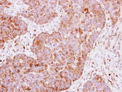
Immunohistochemical analysis of paraffin-embedded human colon carcinoma, using IGF2BP2(GTX113922) antibody at 1:250 dilution.
Antigen Retrieval: Trilogy? (EDTA based, pH 8.0) buffer, 15min
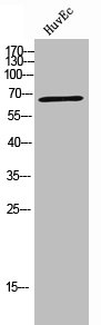
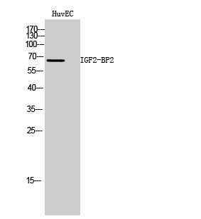
 were separated by 7.5% SDS-PAGE, and the membrane was blotted with IGF2BP2 antibody (GTX113922) diluted at 1:3000. The HRP-conjugated anti-rabbit IgG antibody (GTX213110-01) was used to detect the primary antibody.")
![IGF2BP2 antibody [N1N3] detects IGF2BP2 protein at cytoplasm by immunofluorescent analysis. Sample: HeLa cells were fixed in 4% paraformaldehyde at RT for 15 min. Green: IGF2BP2 protein stained by IGF2BP2 antibody [N1N3] (GTX113922) diluted at 1:500. Blue: Hoechst 33343 staining.](https://www.genetex.com/upload/website/prouct_img/normal/GTX113922/GTX113922_40150_IFA_w_23060501_996.webp "IGF2BP2 antibody [N1N3] detects IGF2BP2 protein at cytoplasm by immunofluorescent analysis. Sample: HeLa cells were fixed in 4% paraformaldehyde at RT for 15 min. Green: IGF2BP2 protein stained by IGF2BP2 antibody [N1N3] (GTX113922) diluted at 1:500. Blue: Hoechst 33343 staining.")
Immunohistochemical analysis of paraffin-embedded human colon carcinoma, using IGF2BP2(GTX113922) antibody at 1:250 dilution.
Antigen Retrieval: Trilogy? (EDTA based, pH 8.0) buffer, 15min
IGF2BP2 antibody
GTX113922
ApplicationsImmunoFluorescence, Western Blot, ImmunoCytoChemistry, ImmunoHistoChemistry, ImmunoHistoChemistry Frozen, ImmunoHistoChemistry Paraffin
Product group Antibodies
ReactivityHuman, Mouse
TargetIGF2BP2
Overview
- SupplierGeneTex
- Product NameIGF2BP2 antibody
- Delivery Days Customer9
- Application Supplier NoteWB: 1:500-1:3000. ICC/IF: 1:100-1:1000. IHC-P: 1:100-1:1000. *Optimal dilutions/concentrations should be determined by the researcher.Not tested in other applications.
- ApplicationsImmunoFluorescence, Western Blot, ImmunoCytoChemistry, ImmunoHistoChemistry, ImmunoHistoChemistry Frozen, ImmunoHistoChemistry Paraffin
- CertificationResearch Use Only
- ClonalityPolyclonal
- Concentration1 mg/ml
- ConjugateUnconjugated
- Gene ID10644
- Target nameIGF2BP2
- Target descriptioninsulin like growth factor 2 mRNA binding protein 2
- Target synonymsIMP-2, IMP2, VICKZ2, insulin-like growth factor 2 mRNA-binding protein 2, IGF-II mRNA-binding protein 2, IGF2 mRNA-binding protein 2, VICKZ family member 2
- HostRabbit
- IsotypeIgG
- Protein IDQ9Y6M1
- Protein NameInsulin-like growth factor 2 mRNA-binding protein 2
- Scientific DescriptionThis gene encodes a member of the IGF-II mRNA-binding protein (IMP) family. The protein encoded by this gene contains several four KH domains and two RRM domains. It functions by binding to the 5 UTR of the insulin-like growth factor 2 (IGF2) mRNA and regulating IGF2 translation. Alternate transcriptional splice variants, encoding different isoforms, have been characterized. [provided by RefSeq]
- ReactivityHuman, Mouse
- Storage Instruction-20°C or -80°C,2°C to 8°C
- UNSPSC41116161
Datasheet
Related products
Product group Antibodies
IGF2BP2 AntibodyCSB-PA006676
ApplicationsWestern Blot, ELISA
ReactivityHuman
TargetIGF2BP2
- SizePrice
Product group Antibodies
Anti-IGF2BP2 AntibodyA100160
ApplicationsWestern Blot, ELISA
ReactivityHuman
- SizePrice

Product group Antibodies
ApplicationsImmunoFluorescence, Western Blot, ImmunoCytoChemistry, ImmunoHistoChemistry
ReactivityHuman, Mouse, Rat
TargetIGF2BP2
- SizePrice
Product group Antibodies

Goat anti-IGF2BP2EB08289
ApplicationsWestern Blot, ELISA
ReactivityCanine, Human, Mouse
TargetIGF2BP2
- SizePrice

Product group Antibodies
Anti-IGF2BP2 AntibodyHPA035145
ApplicationsImmunoCytoChemistry, ImmunoHistoChemistry
ReactivityHuman
TargetIGF2BP2
- SizePrice
Product group Antibodies

IGF2BP2 Antibody (Preservative Free)LS-C343122
ApplicationsWestern Blot, ELISA
ReactivityHuman
TargetIGF2BP2
- SizePrice
Product group Antibodies
ApplicationsImmunoPrecipitation, Western Blot, ImmunoCytoChemistry, ImmunoHistoChemistry
TargetIGF2BP2
- SizePrice
![FACS analysis of HEK293T cells transfected with either IGF2BP2 plasmid(Red) or empty vector control plasmid(Blue) using GTX84303 IGF2BP2 antibody [3G7].](https://www.genetex.com/upload/website/prouct_img/normal/GTX84303/GTX84303_349_FACS_w_23061420_558.webp)
Product group Antibodies
IGF2BP2 antibody [3G7]GTX84303
ApplicationsFlow Cytometry, ImmunoFluorescence, Western Blot, ImmunoCytoChemistry, ImmunoHistoChemistry, ImmunoHistoChemistry Paraffin
ReactivityCanine, Human, Monkey
TargetIGF2BP2
- SizePrice
![IHC-P analysis of human kidney tissue using GTX84306 IGF2BP2 antibody [3F9]. Antigen retrieval : Heat-induced epitope retrieval by 10mM citrate buffer, pH6.0, 100oC for 10min. Dilution : 1:50](https://www.genetex.com/upload/website/prouct_img/normal/GTX84306/GTX84306_2586_IHC-P_w_23061420_904.webp)
Product group Antibodies
IGF2BP2 antibody [3F9]GTX84306
ApplicationsFlow Cytometry, ImmunoFluorescence, Western Blot, ImmunoCytoChemistry, ImmunoHistoChemistry, ImmunoHistoChemistry Paraffin
ReactivityCanine, Human, Monkey, Rat
TargetIGF2BP2
- SizePrice