Bio-Connect
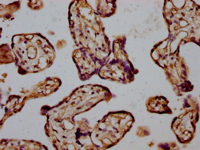
Immunohistochemical staining of human stomach shows moderate to strong cytoplasmic positivity in glandular cells.

Anti-INF2 Antibody

HPA000724
Atlas Antibodies
ApplicationsImmunoHistoChemistry
Product group Antibodies
ReactivityHuman
TargetINF2
Sign in to order and to see your custom pricing.
Large volume orders?
Order with a bulk request

Overview

  • Supplier
    Atlas Antibodies
  • Product Name
    Anti-INF2
  • Delivery Days Customer
    4
  • Applications
    ImmunoHistoChemistry
  • Certification
    Research Use Only
  • Clonality
    Polyclonal
  • Conjugate
    Unconjugated
  • Gene ID64423
  • Target name
    INF2
  • Target description
    inverted formin 2
  • Target synonyms
    C14orf151; C14orf173; CMTDIE; FSGS5; HBEAG-binding protein 2 binding protein C; HBEBP2-binding protein C; inverted formin, FH2 and WH2 domain containing; inverted formin-2; pp9484
  • Host
    Rabbit
  • Isotype
    IgG
  • Protein IDQ27J81
  • Protein Name
    Inverted formin-2
  • Scientific Description
    Recombinant Protein Epitope Signature Tag (PrEST) antigen sequence
  • Reactivity
    Human
  • Storage Instruction
    -20°C,2°C to 8°C
  • UNSPSC
    41116161

Related products

Product group Antibodies
Bethyl Laboratories
ApplicationsImmunoPrecipitation, Western Blot
TargetINF2
  • SizePrice
IHC image of CSB-PA637740LA01HU diluted at 1:400 and staining in paraffin-embedded human placenta tissue performed on a Leica BondTM system. After dewaxing and hydration, antigen retrieval was mediated by high pressure in a citrate buffer (pH 6.0). Section was blocked with 10% normal goat serum 30min at RT. Then primary antibody (1% BSA) was incubated at 4°C overnight. The primary is detected by a biotinylated secondary antibody and visualized using an HRP conjugated SP system.
Product group Antibodies
Cusabio
INF2 AntibodyCSB-PA637740LA01HU
ApplicationsELISA, ImmunoHistoChemistry
ReactivityHuman
TargetINF2
  • SizePrice
Atlas Antibodies
Protein IDQ27J81
  • SizePrice
Figure 1. Western blot analysis of INF2 using anti-INF2 antibody (A02710-1). Electrophoresis was performed on a 5-20% SDS-PAGE gel at 70V (Stacking gel) / 90V (Resolving gel) for 2-3 hours. The sample well of each lane was loaded with 30 ug of sample under reducing conditions. Lane 1: human Hela whole cell lysates, Lane 2: human A549 whole cell lysates, Lane 3: rat C6 whole cell lysates, Lane 4: mouse brain tissue lysates. After electrophoresis, proteins were transferred to a nitrocellulose membrane at 150 mA for 50-90 minutes. Blocked the membrane with 5% non-fat milk/TBS for 1.5 hour at RT. The membrane was incubated with rabbit anti-INF2 antigen affinity purified polyclonal antibody (Catalog # A02710-1) at 0.5 microg/mL overnight at 4°C, then washed with TBS-0.1%Tween 3 times with 5 minutes each and probed with a goat anti-rabbit IgG-HRP secondary antibody at a dilution of 1:5000 for 1.5 hour at RT. The signal is developed using an Enhanced Chemiluminescent detection (ECL) kit (Catalog # EK1002) with Tanon 5200 system. A specific band was detected for INF2 at approximately 180 kDa. The expected band size for INF2 is at 136 kDa.
Product group Antibodies
Boster Bio
Anti-INF2 Antibody Picoband(r)A02710-1-CARRIER-FREE
ApplicationsFlow Cytometry, ImmunoFluorescence, ImmunoPrecipitation, Western Blot, ELISA, ImmunoCytoChemistry
TargetINF2
  • SizePrice
Product group Antibodies
GeneTex
INF2 antibodyGTX130714
ApplicationsImmunoPrecipitation, Western Blot
TargetINF2
  • SizePrice
Product group Antibodies
Applied Biological Materials
Anti-INF2Y058773
ApplicationsWestern Blot, ELISA, ImmunoHistoChemistry
  • SizePrice
Product group Antibodies
LifeSpan BioSciences
INF2 AntibodyLS-C834903
ApplicationsELISA, ImmunoHistoChemistry
TargetINF2
  • SizePrice